

A method for measuring brain partial pressure of oxygen in unanesthetized unrestrained subjects: the effect of acute and chronic hypoxia on brain tissue PO(2)
- PMID: 20817029
- PMCID: PMC3044503
- DOI: 10.1016/j.jneumeth.2010.08.019
A method for measuring brain partial pressure of oxygen in unanesthetized unrestrained subjects: the effect of acute and chronic hypoxia on brain tissue PO(2)
Erratum in
- J Neurosci Methods. 2011 Mar 30;196(2):327
Abstract
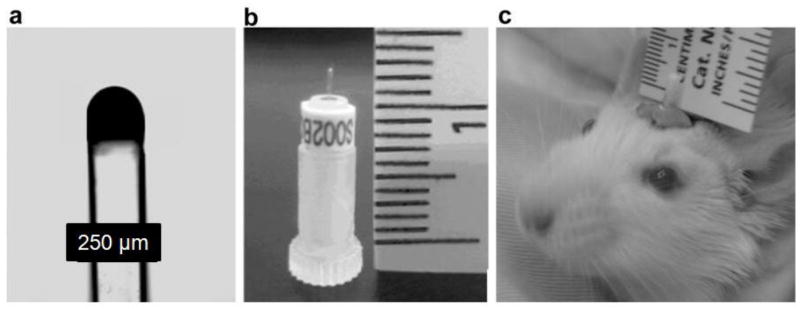
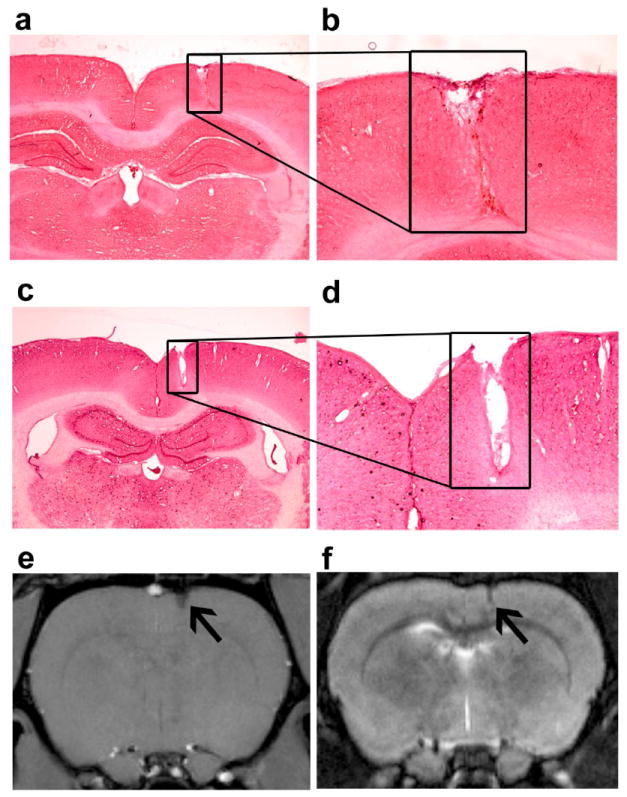
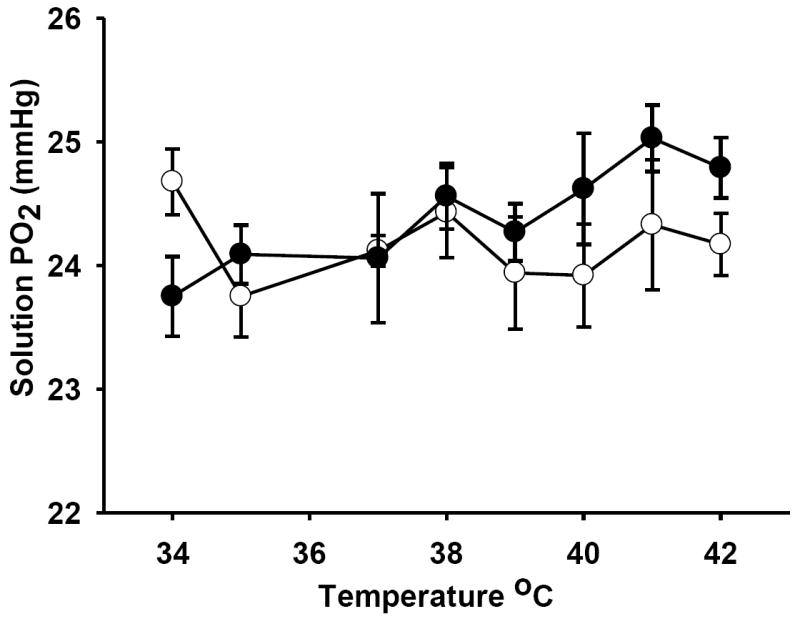
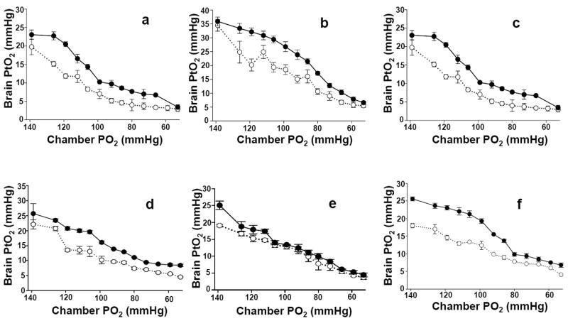
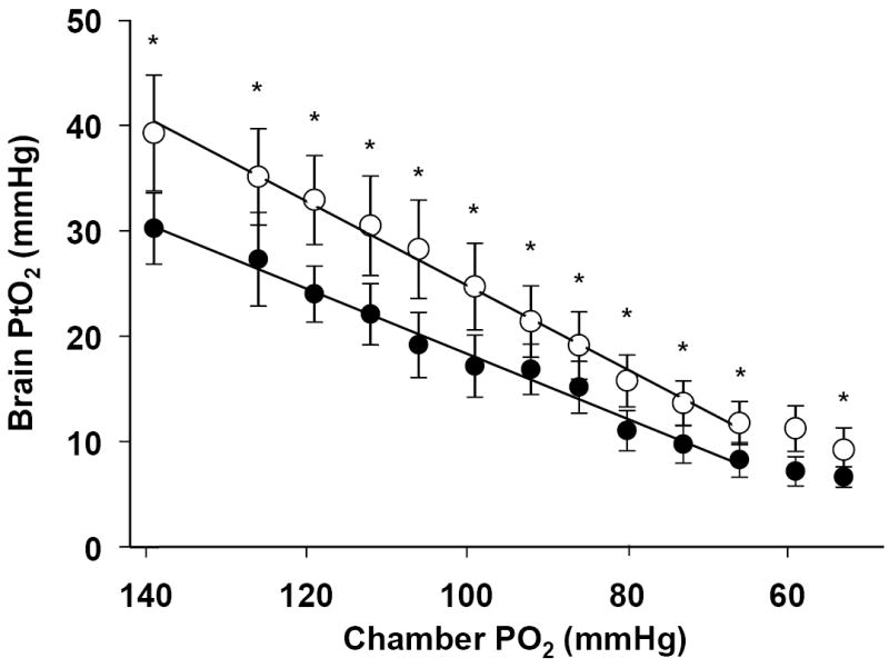
The level of tissue oxygenation provides information related to the balance between oxygen delivery, oxygen utilization, tissue reactivity and morphology during physiological conditions. Tissue partial pressure of oxygen (PtO(2)) is influenced by the use of anesthesia or restraint. These factors may impact the absolute level of PtO(2). In this study we present a novel fiber optic method to measure brain PtO(2). This method can be used in unanesthetized, unrestrained animals, provides absolute values for PO(2), has a stable calibration, does not consume oxygen and is MRI compatible. Brain PtO(2) was studied during acute hypoxia, as well as before and after 28 days of high altitude acclimatization. A sensor was chronically implanted in the frontal cortex of eight Wistar rats. It is comprised of a fiber optic probe with a tip containing material that fluoresces with an oxygen dependent lifetime. Brain PtO(2) declines by 80% and 76% pre- and post-acclimatization, respectively, when the fraction of inspired oxygen declines from 0.21 to 0.08. In addition, a linear relationship between brain PtO(2) and inspired O(2) levels was demonstrated r(2)=0.98 and r(2)=0.99 (pre- and post-acclimatization). Hypoxia acclimatization resulted in an increase in the overall brain PtO(2) by approximately 35%. This paper demonstrates the use of a novel chronically implanted fiber optic based sensor for measuring absolute PtO(2). It shows a very strong linear relationship in awake animals between inspired O(2) and tissue O(2), and shows that there is a proportional increase in PtO(2) over a range of inspired values after exposure to chronic hypoxia.
Copyright © 2010 Elsevier B.V. All rights reserved.
Figures
References
-
- Bardt TF, Unterberg AW, Hartl R, Kiening KL, Schneider GH, Lanksch WR. Monitoring of brain tissue PO2 in traumatic brain injury: effect of cerebral hypoxia on outcome. Acta Neurochirurgica - Supplementum. 1998;71:153–6. - PubMed
-
- Bazzu G, Puggioni GG, Dedola S, Calia G, Rocchitta G, Migheli R, Desole MS, Lowry JP, O’Neill RD, Serra PA. Real-Time Monitoring of Brain Tissue Oxygen Using a Miniaturized Biotelemetric Device Implanted in Freely Moving Rats. Anal Chem. 2009;81:2235–41. - PubMed
-
- Beck T, Krieglstein J. Cerebral circulation, metabolism, and blood-brain barrier of rats in hypocapnic hypoxia. Am J Physiol. 1987;252:H504–12. - PubMed
-
- Boero JA, Ascher J, Arregui A, Rovainen C, Woolsey TA. Increased brain capillaries in chronic hypoxia. J Appl Physiol. 1999;86:1211–9. - PubMed
-
- Borgstrom L, Johannsson H, Siesjo BK. The relationship between arterial PO2 and cerebral blood flow in hypoxic hypoxia. Acta Physiol Scand. 1975;93:423–32. - PubMed
Publication types
MeSH terms
Substances
Grants and funding
LinkOut - more resources
Full Text Sources
