Review
. 2010 Dec;20(12):734-42.
doi: 10.1016/j.tcb.2010.08.002.

Diverse evolutionary paths to cell adhesion

Affiliations
Review

Diverse evolutionary paths to cell adhesion

Monika Abedin et al. Trends Cell Biol. 2010 Dec.

Abstract

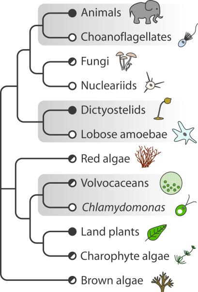
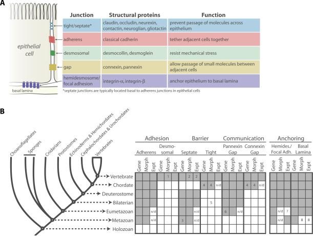
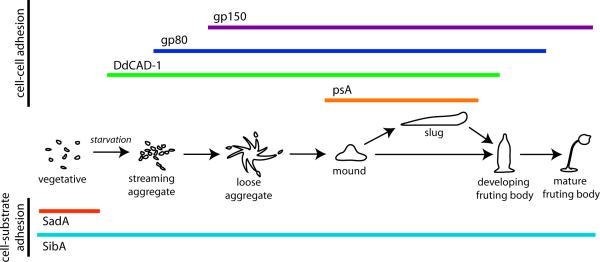
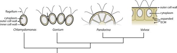
The morphological diversity of animals, fungi, plants, and other multicellular organisms stems from the fact that each lineage acquired multicellularity independently. A prerequisite for each origin of multicellularity was the evolution of mechanisms for stable cell-cell adhesion or attachment. Recent advances in comparative genomics and phylogenetics provide critical insights into the evolutionary foundations of cell adhesion. Reconstructing the evolution of cell junction proteins in animals and their unicellular relatives exemplifies the roles of co-option and innovation. Comparative studies of volvocine algae reveal specific molecular changes that accompanied the evolution of multicellularity in Volvox. Comparisons between animals and Dictyostelium show how commonalities and differences in the biology of unicellular ancestors influenced the evolution of adhesive mechanisms. Understanding the unicellular ancestry of cell adhesion helps illuminate the basic cell biology of multicellular development in modern organisms.


Figures

Figure 1
Figure 1. Diverse multicellular eukaryotes and their closest unicellular or colonial relatives
The phylogenetic relationships among select unicellular, colonial and multicellular eukaryotic lineages indicate that multicellularity evolved multiple times. Some lineages are strictly multicellular (filled circle) and some are unicellular or display simple/undifferentiated colonies (open circle), while others have a mix of unicellular or colonial and multicellular forms (half filled circle). Comparisons among multicellular lineages and their closest unicellular relatives provide insights into the mechanisms underlying transitions to multicellularity. Lineages discussed in this review are highlighted in grey.
Figure 2
Figure 2. Phylogenetic distribution of epithelial junctions based on ultrastructural, genomic and functional data
A) Schematic representation of animal epithelial cell junctions. Each type of junction, comprised of a unique set of structural proteins, is restricted to a specific region of the epithelial cell membrane and serves a distinct function (indicated in table). (B) Evolutionary history of epithelial cell junctions. The four main functions of animal epithelia are adhesion, barrier, signaling and anchoring. Elements of adherens, septate, pannexin-based gap and anchoring junctions [hemidesmosomes (Hemides.), focal adhesion (Focal Adh.) and the basal lamina] were likely present in the last common ancestor of animals while desmosomes, tight and connexin-based gap junctions emerged later in animal evolution. For a series of ancestors representing stages in animal evolution, the presence (filled box) or absence (open box) of evidence for (1) genes diagnostic of each junction type (“Gene”), (2) junction morphology detected by electron microscopy (“Morph”) and (3) experimental support (e.g. stable cell adhesion, barrier function, or protein localization; “Expt”) in extant lineages is indicated [, –, , –38, 70, 71]. Numbered boxes indicate observations of note: 1, reports of desmosomes observed in non-vertebrates by electron microscopy are controversial [11]; 2, septate junctions have been identified in mammals but not in other chordates [16]; 3, evidence for septate junctional proteins in sponges is based solely on BLAST analysis of EST data [19]; 4, tight and connexin-based gap junctions are found in Urochordates but not Cephalochordates [, –28]; 5, a single example of tight junctions has been reported in the spider central nervous system [72]; 6, pannexins are present in Hydra but absent from the Nematostella genome suggesting that they are not found in all cnidarians [23, 25]; 7, while cell biological and biochemical investigations of integrin function in cnidaria is absent, preliminary research in the jellyfish Podocoryne carnea indicates that transcripts of integrin-α and -β subunits have been detected in the same cells as talin, a focal adhesion protein [73]; 8, although all sponges have ECM, only some sponges display structures that resemble basal lamina and only homoscleromorph sponges have been reported to possess ECM comprised of type-IV collagen, a major component of the basal lamina [–37]; n/d, not determined.
Figure 3
Figure 3. Cell adhesion proteins are dynamically expressed during D. discoideum development
Upon starvation, a complex developmental program is initiated involving temporal regulation of diverse cell-cell and cell-substrate adhesion proteins. Colored bars indicate timing of gene expression. The cell-cell adhesion protein DdCAD-1 is expressed when aggregation initiates and is followed by gp80 and then gp150. PsA expression is initiated at the mound stage and persists until fruiting body development. The cell-substrate adhesion protein SadA is active only during vegetative growth while SibA is constitutively expressed. Adapted from [46].
Figure 4
Figure 4. Evolution of cell wall into ECM in the volvocine algae
The graded complexity of volvocine algae coupled with insights into their phylogenetic relationships provide the opportunity to identify the molecular foundations of an evolutionary transition to multicellularity. Chlamydomonas possesses an HRGP rich cell wall consisting of an outer cell wall (dark grey) with a characteristic tripartite structure (three fibrous layers, termed the `tripartite layer') and a relatively amorphous inner cell wall (brown) surrounding the cell membrane and cytoplasm. Cells in Gonium colonies have similar cell walls that are attached to neighboring walls by ECM-based structures (arrow). In Pandorina, the outer cell wall surrounds the entire colony and the cells are embedded in a common ECM comprised of inner cell wall HRGP homologs. Multicellular Volvox has a voluminous ECM derived from diverse HRGP family members that have specialized functions. Adapted from [56].
box 1
box 1

References

    1. Jarvis MC, et al. Intercellular adhesion and cell separation in plants. Plant Cell Environ. 2003;26:977–989.
    1. Gumbiner BM. Cell adhesion: the molecular basis of tissue architecture and morphogenesis. Cell. 1996;84:345–357. - PubMed
    1. King N. The unicellular ancestry of animal development. Dev. Cell. 2004;7:313–325. - PubMed
    1. Baldauf SL. The deep roots of eukaryotes. Science. 2003;300:1703–1706. - PubMed
    1. Carroll SB. Chance and necessity: the evolution of morphological complexity and diversity. Nature. 2001;409:1102–1109. - PubMed

Publication types