. 2010 Nov 19;285(47):36897-908.
doi: 10.1074/jbc.M110.174391. Epub 2010 Sep 3.

Sequence-dependent prion protein misfolding and neurotoxicity

Affiliations

Sequence-dependent prion protein misfolding and neurotoxicity

Pedro Fernandez-Funez et al. J Biol Chem. .

Abstract

Prion diseases are neurodegenerative disorders caused by misfolding of the normal prion protein (PrP) into a pathogenic "scrapie" conformation. To better understand the cellular and molecular mechanisms that govern the conformational changes (conversion) of PrP, we compared the dynamics of PrP from mammals susceptible (hamster and mouse) and resistant (rabbit) to prion diseases in transgenic flies. We recently showed that hamster PrP induces spongiform degeneration and accumulates into highly aggregated, scrapie-like conformers in transgenic flies. We show now that rabbit PrP does not induce spongiform degeneration and does not convert into scrapie-like conformers. Surprisingly, mouse PrP induces weak neurodegeneration and accumulates small amounts of scrapie-like conformers. Thus, the expression of three highly conserved mammalian prion proteins in transgenic flies uncovered prominent differences in their conformational dynamics. How these properties are encoded in the amino acid sequence remains to be elucidated.


Figures

FIGURE 1.
FIGURE 1.
Sequence alignment and expression of hamster, mouse, and rabbit PrP in flies. A, ClustalW alignment of the globular domain of hamster (residues 122–231), mouse (residues 121–230), and rabbit (residues 123–231) PrP. The β-sheet domains (blue) and α-helices (red) are indicated. Color coding of amino acids is as follows: red, small and/or hydrophobic; blue, acidic; magenta, basic; green, hydrophilic, charged. L42 indicates a RaPrP-specific epitope recognized by the L42 antibody that has no effect when introduced in MoPrP. The red box in MoV214 indicates a position key for MoPrP conversion with a conservative change in RaPrP and a radical change in HaPrP. B, detection of hamster, mouse, and rabbit PrP expressed in transgenic flies by Western blot. A mix of 3F4 and 6H4 antibodies was used for detection of the three proteins in the same membrane. HaPrP and MoPrP accumulate in two bands, whereas RaPrP accumulates in three, the top two being the more prominent. C, relative expression of PrP transcripts by quantitative RT-PCR. Two HaPrP lines, moderate (M9) and strong (M6), are shown as reference. The MoPrP-P1 line induces slightly lower (nonsignificant) levels than the strong HaPrP-M6 line. The RaPrP-F22 line induces 20% more PrP transcripts than HaPrP-M6. Values were normalized with RNA polymerase II transcript levels.
FIGURE 2.
FIGURE 2.
Spongiform degeneration of brain neurons. A and B, brain morphology of a 1-day-old fly expressing HaPrP (da-Gal4/HaPrP). A, at low magnification, the optic lobes (OL) and the central brain (Br) exhibit well preserved anatomy. B, in more detail (box in A), the cortex (CO) contains a thick layer of cell bodies, and the neuropile fills all the space below. C and D, spongiform degeneration in 30-day-old flies expressing HaPrP. C, these brains are very thin and contain abundant vacuoles in the central brain (red arrow) and the optic lobes (white arrows). D, at higher magnification, the neuropile contains large vacuoles (red arrows), whereas the cortex is very thin and contains multiple microvacuoles (arrowheads). E and F, brain of 30-day-old flies expressing MoPrP. E, these brains display normal size and contain few vacuoles, mostly in the optic lobes (white arrows). F, at higher magnification, the cortex shows few microvacuoles and a larger cortex that contains more cells than the HaPrP flies. G and H, brain of 30-day-old flies expressing RaPrP. G, RaPrP brains are very similar to MoPrP; they are large and contain few vacuoles, typically in the optic lobes (white arrow). H, neuropile contains no vacuoles, whereas cortex is larger and shows few microvacuoles (arrowhead).
FIGURE 3.
FIGURE 3.
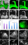
Only HaPrP induces degeneration of the mushroom bodies. A, three-dimensional reconstruction of a Z-stack displaying the mushroom bodies (MB) in front and the Kenyon cells (Kc) in the posterior brain of a 1-day-old control fly (OK107-Gal4/CD8-GFP). d, dorsal; med, medial; a, anterior; p, posterior. B–F, mushroom body degeneration. B, mushroom bodies contain three lobes, α, β, and γ, which exhibit normal morphology in 40-day-old control flies (OK107-Gal4/CD8-GFP/LacZ; GFP shown). C, mushroom bodies display normal morphology in 1-day-old flies expressing HaPrP (OK107-Gal4/CD8-GFP/HaPrP; GFP shown). D, in 40-day-old flies expressing HaPrP, the distal portion of the α lobe is missing (arrow, HaPrP shown). The inset shows axonal blebbing of the retracting α lobe (arrowheads, GFP shown). E and F, α lobe (arrow) appears normal in 40-day-old flies expressing MoPrP (OK107-Gal4/CD8-GFP/MoPrP; MoPrP shown) or RaPrP (OK107-Gal4/CD8-GFP/RaPrP; RaPrP shown). The thicker aspect of E is due to higher antibody signal. G–K, degeneration of Kenyon cells. G, young control flies (genotype as in B, n = 6) and flies expressing HaPrP (genotype as in C, n = 7) contain approximately the same number of Kenyon cells as estimated by the surface they occupy. These clusters are larger in 40-day-old control flies (GFP shown, n = 10) (H). 40-Day-old flies expressing HaPrP show a significant reduction in the number of Kenyon cells (I, HaPrP shown, n = 8). 40-Day-old flies expressing MoPrP (J, MoPrP shown, n = 19) or RaPrP (K, RaPrP shown, n = 14) present a modest reduction in the size of the clusters, although only the MoPrP were significantly smaller. Values in H–K indicate % surface occupied by Kc clusters compared with controls (H). L–N, high magnification images of Kenyon cells in the edge of the clusters. L, HaPrP accumulates in distinct puncta (arrows). M, MoPrP has a mixed punctate (arrow) and diffuse expression, and N, RaPrP is mostly diffuse (arrows).
FIGURE 4.
FIGURE 4.
Subcellular distribution of PrP in Drosophila neurons. A–D, high magnification confocal images of motor neurons expressing PrP (BG380-Gal4/UAS-PrP). A, in permeabilized samples, HaPrP (magenta) accumulates in cytoplasmic puncta (arrows), although the extracellular neuronal marker HRP (green) weakly outlines the neuronal contour. B, in the absence of detergent (−Triton), both HaPrP and HRP label the surface of the cell body. C, MoPrP accumulates in a few puncta (arrows), and some protein labels diffusely the rest of the cell (arrowhead). D, RaPrP shows diffuse perinuclear accumulation, suggesting localization in the ER (arrowhead). E–I, high magnification confocal images of interneurons in the ventral cord expressing PrP under the control of OK107-Gal4. E, HaPrP (magenta) accumulates in distinct cytosolic puncta that co-localize with the Golgi marker galactosyltransferase (GalT-GFP, green, arrows); F, Golgi-secretory vesicle reporter Rab11-GFP (green, arrows); G, but not with mitochondria (mito-GFP, green). H, MoPrP (magenta) accumulates in distinct cytosolic puncta that co-localize with Rab11-GFP (green, arrows) and diffusely in the ER (arrowhead). I, RaPrP (magenta) accumulates with a diffuse distribution in the ER (arrowhead) and the membrane by co-localization with KDEL-GFP (green).
FIGURE 5.
FIGURE 5.
Hamster and mouse PrP induce locomotor dysfunction. A, climbing ability of adult males expressing LacZ (control, blue), HaPrP (red), MoPrP (green), and RaPrP (orange) in motor neurons (BG380-Gal4). Control flies move well for 26 days (over 50% climbing ability) and preserve some activity when the experiment was stopped after 31 days. Hamster and mouse PrP induce early locomotor dysfunction (days 3 and 4, respectively), although RaPrP induces a milder locomotor phenotype (day 21) that almost parallels the control flies. B–E, software-assisted video analysis of fly locomotion. B, after creating custom arenas that cover the vials, the software can identify and track flies inside the arena for 20 s. C, analysis of active flies (n = 3). HaPrP flies are less active than Mo- and RaPrP. D, analysis of average speed of active flies (n = 3). HaPrP flies are significantly slower than Mo- and RaPrP. E, total distance traveled by active flies (n = 3). HaPrP flies cover significantly less distance than Mo- and RaPrP.
FIGURE 6.
FIGURE 6.
Differential biochemical properties of hamster, mouse, and rabbit PrP in flies. A, PrP insolubility. Flies expressing Ha-, Mo-, or RaPrP ubiquitously were homogenized at days 1 and 30 and assayed for solubility in 10% Sarkosyl/NaPTA. The soluble (S) and insoluble (I) fractions were loaded in a gel together with an equivalent volume of the total (T) homogenate. All three prion proteins accumulate mostly in the soluble fraction in younger flies. In the older flies, a significant fraction of PrP accumulates in the insoluble fraction. HaPrP seems to have the largest amount of insoluble PrP, although RaPrP seems to accumulate the lowest amount. The bottom row shows the distribution of α-tubulin as a quality control for the procedure. B, acquisition of PrPSc-like conformations. Homogenates from flies expressing Ha-, Mo-, or RaPrP ubiquitously (genotypes as in A) were incubated with 15B3-coated beads (+) or control beads (−). 1-Day-old flies expressing HaPrP produced very low reactivity against 15B3, but 30-day-old flies showed strong signal. MoPrP flies also showed very weak signal in young flies and stronger immunoreactivity in older flies (weaker than HaPrP). Neither young nor old RaPrP flies rendered signal. Beads without the 15B3 antibody produced no signal in the older flies for each construct. C–E, accumulation of large PrP aggregates. Tissue lysates from young and old flies (same as in A) were loaded on top of sucrose gradients and centrifuged, and 12 fractions were collected, from low (top = 1) to high molecular weight (bottom = 12). In young flies expressing hamster, mouse, or rabbit PrP (top panels), most PrP accumulated in the low density fractions (1 and 2). Young flies expressing HaPrP accumulate small amounts of PrP in fractions 10–12 (C, top panel), although older flies accumulated large amounts (C, bottom panel). Flies expressing MoPrP also accumulated PrP in fractions 10–12 but much less than the HaPrP flies (D, bottom panel). Flies expressing RaPrP only accumulated trace amounts of PrP in the high molecular weight fractions (E, bottom panel).
FIGURE 7.
FIGURE 7.
Characterization of aggregates size for three mammalian PrP. A–C, determination of the size of PrP aggregates in tissue homogenates from flies expressing Ha-, Mo-, or RaPrP ubiquitously (da-Gal/UAS-PrP) by size exclusion chromatography. 100 fractions were collected, and the presence of PrP was examined by Western blot. Molecular weight was determined with the use of appropriate markers. A–C, top panels, in young flies, most of the PrP is found in fractions 60–66 with a molecular mass of 66 kDa or less. These PrP molecules are either monomeric or form small oligomers. In the young flies, only HaPrP is present in fractions 30–34 with a molecular mass of more than 2,000 kDa. A–C, bottom panels, in older flies, PrP accumulates with a slight left shift of the smaller particles (fractions 58–62), suggesting an increase in the amount and size of oligomers and a reduction of monomers. In the older flies, HaPrP accumulates in high amounts in fractions 30–34. MoPrP and RaPrP only accumulate in trace amounts in these fractions.
FIGURE 8.
FIGURE 8.
Conformational transitions of mammalian PrP in flies. Transgenic flies expressing hamster, mouse, and rabbit PrP accumulate different conformers based on their biochemical and structural properties. The three proteins accumulate initially with properties typical of PrPC. However, older flies accumulate Sarkosyl-insoluble PrP, consistent with PrP misfolding. RaPrP may accumulate in a unique misfolded conformation that is not 15B3 immunoreactive and that we called PrPinsol. Both HaPrP and MoPrP produce 15B3 epitopes, indicating that they accumulate PrPSc-like or PrP* isoforms. However, only HaPrP accumulated large amounts of high molecular weight aggregates, suggesting that this is also a unique conformer (PrPaggre). Some of these isoforms correlate with specific degenerative phenotypes, and PrPaggre is the only conformer associated with spongiform degeneration.

References

    1. Aguzzi A., Heikenwalder M., Polymenidou M. (2007) Nat. Rev. Mol. Cell Biol. 8, 552–561 - PubMed
    1. Barlow R. M., Rennie J. C. (1976) Res. Vet. Sci. 21, 110–111 - PubMed
    1. Gibbs C. J., Jr., Gajdusek D. C. (1973) Science 182, 67–68 - PubMed
    1. Courageot M. P., Daude N., Nonno R., Paquet S., Di Bari M. A., Le Dur A., Chapuis J., Hill A. F., Agrimi U., Laude H., Vilette D. (2008) J. Gen. Virol. 89, 341–347 - PubMed
    1. Vilette D., Andreoletti O., Archer F., Madelaine M. F., Vilotte J. L., Lehmann S., Laude H. (2001) Proc. Natl. Acad. Sci. U.S.A. 98, 4055–4059 - PMC - PubMed

Publication types

MeSH terms

LinkOut - more resources