. 2010 Nov 5;285(45):34972-80.
doi: 10.1074/jbc.M110.141838. Epub 2010 Sep 3.

The exquisite structure and reaction mechanism of bacterial Pz-peptidase A toward collagenous peptides: X-ray crystallographic structure analysis of PZ-peptidase a reveals differences from mammalian thimet oligopeptidase

Affiliations

The exquisite structure and reaction mechanism of bacterial Pz-peptidase A toward collagenous peptides: X-ray crystallographic structure analysis of PZ-peptidase a reveals differences from mammalian thimet oligopeptidase

Akio Kawasaki et al. J Biol Chem. .

Abstract

Pz-peptidase A, from the thermophilic bacterium Geobacillus collagenovorans MO-1, hydrolyzes a synthetic peptide substrate, 4-phenylazobenzyloxycarbonyl-Pro-Leu-Gly-Pro-D-Arg (Pz-PLGPR), which contains a collagen-specific tripeptide sequence, -Gly-Pro-X-, but does not act on collagen proteins themselves. The mammalian enzyme, thimet oligopeptidase (TOP), which has comparable functions with bacterial Pz-peptidases but limited identity at the primary sequence level, has recently been subjected to x-ray crystallographic analysis; however, no crystal structure has yet been reported for complexes of TOP with substrate analogues. Here, we report crystallization of recombinant Pz-peptidase A in complex with two phosphinic peptide inhibitors (PPIs) that also function as inhibitors of TOP and determination of the crystal structure of these complexes at 1.80-2.00 Å resolution. The most striking difference between Pz-peptidase A and TOP is that there is no channel running the length of bacterial protein. Whereas the structure of TOP resembles an open bivalve, that of Pz-peptidase A is closed and globular. This suggests that collagenous peptide substrates enter the tunnel at the top gateway of the closed Pz-peptidase A molecule, and reactant peptides are released from the bottom gateway after cleavage at the active site located in the center of the tunnel. One of the two PPIs, PPI-2, which contains the collagen-specific sequence, helped to clarify the exquisite structure and reaction mechanism of Pz-peptidase A toward collagenous peptides. This study describes the mode of substrate binding and its implication for the mammalian enzymes.


Figures

FIGURE 1.
FIGURE 1.
Overall structure of Pz-peptidase A with inhibitor PPI-1. A and B, schematic representation of the structure in two different directions. The directions in A and B are perpendicular to each other. These figures, as well as Figs. 2–7, except for Fig. 3, were prepared with the PyMOL program. Twenty five α helices and five β strands are depicted in blue and red, respectively, whereas the loops are in green. The inhibitor PPI-1 molecule, drawn in a stick representation, is in pink. The gray arrow is a direction for Fig. 2.
FIGURE 2.
FIGURE 2.
Comparison of Pz-peptidase A with TOP in light of channel. The overall structures of Pz-peptidase A and TOP are depicted in the same directions as those shown in Fig. 1 with a gray arrow. The space of the deep and narrow channel that runs the length of the TOP molecule and its corresponding one in Pz-peptidase A is indicated with red ovals.
FIGURE 3.
FIGURE 3.
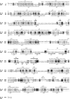
Secondary structure alignment of Pz-peptidase A and human TOP. Conserved sequences are highlighted in black, and semiconservative ones are boxed. Deleted sequences are shown with dots. Secondary structure elements are depicted schematically (coil, α helix; arrow, β strand) above for Pz-peptidase A and below for human TOP.
FIGURE 4.
FIGURE 4.
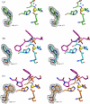
Active site residues of Pz-peptidase A. The active site residues (His356, Glu357, His360, and Glu384) are depicted as sticks, whereas two water molecules and a zinc atom are shown as red and gray balls, respectively. The electron densities corresponding to the active site residues are shown. The map was calculated with coefficients of the form 2FoFc. The map was contoured at 1.0σ.
FIGURE 5.
FIGURE 5.
Tunnel exhibition. Gateways leading to the tunnel (red) for substrates and products were identified using the CAVER program (43) and are represented using the PyMOL program. The direction for the Pz-peptidase A molecule is the same as that for Fig. 1.
FIGURE 6.
FIGURE 6.
Close-up views of inhibitor PPI-2 in the active site. Electron density maps for PPI-2 are depicted. The 2FoFc maps contoured at the 1.0σ level are superposed on stick models of the bound substrates. The main chain of the active site residues (His356, Glu357, His360, and Glu384) are in orange, whereas other residues binding with PPI-2 by H-bonds (Cys329, His479, Tyr486, Tyr487, and Tyr490) are in blue. H-bonds are shown in orange.
FIGURE 7.
FIGURE 7.
Movement of the side chain of Trp388 in the absence (A) and presence of inhibitors PPI-1 (B) or PPI-2 (C). The residue Trp377 is depicted with the 2FoFc maps contoured at the 1.0σ level. The residue Trp377 and the active site residues (His356, Glu357, His360, and Glu384) are shown in green (A), light blue (B), and orange (C), whereas the inhibitors are in pink and a zinc atom is in yellow.

References

    1. Nimni M. E. (1983) Semin. Arthritis Rheum. 13, 1–86 - PubMed
    1. Suzuki Y., Tsujimoto Y., Matsui H., Watanabe K. (2006) J. Biosci. Bioeng. 102, 73–81 - PubMed
    1. Linsenmayer T. F. (1991) in Cell Biology of Extracellular Matrix (Hay E. D. ed.) pp. 7–44, Plenum Press, New York
    1. Myers L. K., Tang B., Rosloniec E. F., Stuart J. M., Chiang T. M., Kang A. H. (1998) J. Immunol. 161, 3589–3595 - PubMed
    1. Harrington D. J. (1996) Infect. Immun. 64, 1885–1891 - PMC - PubMed

Publication types

LinkOut - more resources