. 2010 Sep 21;107(38):16661-6.
doi: 10.1073/pnas.1007397107. Epub 2010 Sep 3.

Axonal α7 nicotinic ACh receptors modulate presynaptic NMDA receptor expression and structural plasticity of glutamatergic presynaptic boutons

Affiliations

Axonal α7 nicotinic ACh receptors modulate presynaptic NMDA receptor expression and structural plasticity of glutamatergic presynaptic boutons

Hong Lin et al. Proc Natl Acad Sci U S A. .

Abstract

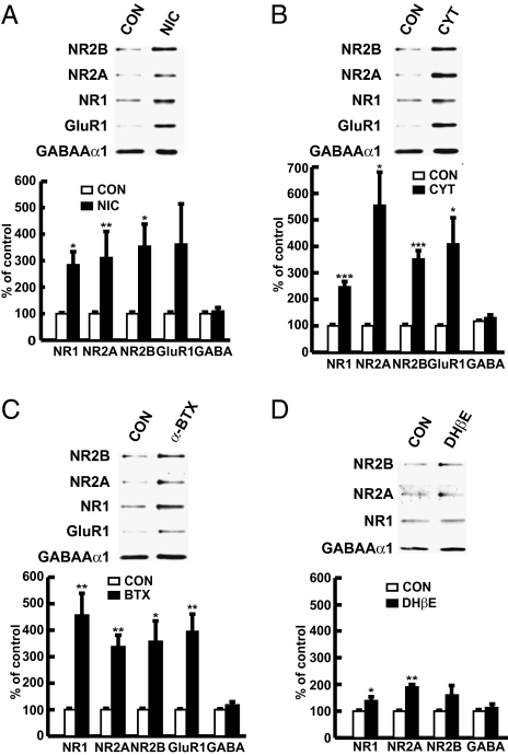
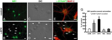
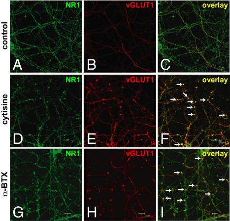
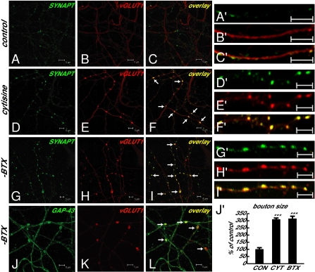
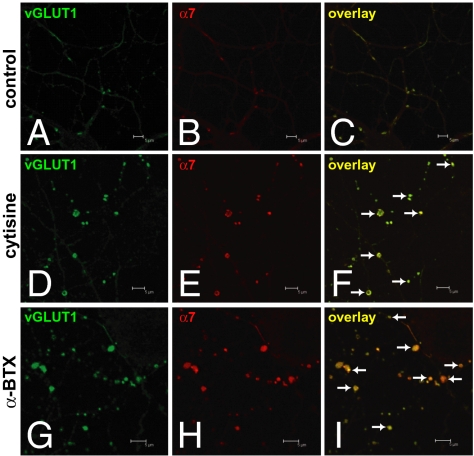
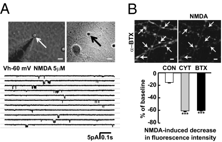
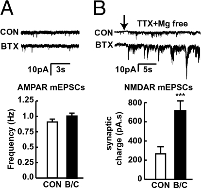
In association with NMDA receptors (NMDARs), neuronal α7 nicotinic ACh receptors (nAChRs) have been implicated in neuronal plasticity as well as neurodevelopmental, neurological, and psychiatric disorders. However, the role of presynaptic NMDARs and their interaction with α7 nAChRs in these physiological and pathophysiological events remains unknown. Here we report that axonal α7 nAChRs modulate presynaptic NMDAR expression and structural plasticity of glutamatergic presynaptic boutons during early synaptic development. Chronic inactivation of α7 nAChRs markedly increased cell surface NMDAR expression as well as the number and size of glutamatergic axonal varicosities in cortical cultures. These boutons contained presynaptic NMDARs and α7 nAChRs, and recordings from outside-out pulled patches of enlarged presynaptic boutons identified functional NMDAR-mediated currents. Multiphoton imaging of presynaptic NMDAR-mediated calcium transients demonstrated significantly larger responses in these enlarged boutons, suggesting enhanced presynaptic NMDAR function that could lead to increased glutamate release. Moreover, whole-cell patch clamp showed a significant increase in synaptic charge mediated by NMDAR miniature EPSCs but no alteration in the frequency of AMPAR miniature EPSCs, suggesting the selective enhancement of postsynaptically silent synapses upon inactivation of α7 nAChRs. Taken together, these findings indicate that axonal α7 nAChRs modulate presynaptic NMDAR expression and presynaptic and postsynaptic maturation of glutamatergic synapses, and implicate presynaptic α7 nAChR/NMDAR interactions in synaptic development and plasticity.


Conflict of interest statement

The authors declare no conflict of interest.

Figures

Fig. 1.
Fig. 1.
Chronic inactivation of α7 nAChR, but not α4β2 nAChR, leads to marked increases in surface level of NR1, NR2, and GluR1 subunits in cortical neurons. Representative blots and quantification showing surface level of NR1, NR2A, NR2B, GluR1, and GABAAα1 in cortical cultures treated with nicotine (A), cytisine (B), α-BTX (C) or DHβE (D). Each experiment was repeated at least three times.
Fig. 2.
Fig. 2.
Chronic inactivation of α7 nAChR increases number and size of NR1-positive axonal varicosities in cortical cultures. Representative images showing increased and enlarged NR1-positive axonal varicosities (B), DIC images of enlarged boutons (D), and α-synaptophysin–positive presynaptic terminals (F) in α-BTX–treated cultures compared with those boutons in control cultures (A, C, and E). Quantification (G) showed that α-BTX, cytisine, but not DHβE or nicotine, increases the number of NR1-positive axonal varicosities in cortical cultures (n = 3–5). (Scale bars, 20 μm.)
Fig. 3.
Fig. 3.
Increased and enlarged NR1-positive axonal varicosities are glutamatergic presynaptic boutons. (A–C) NR1 and vGLUT1 immunoreactivities in control cultures. (D–I) Colocalization of glutamatergic presynaptic terminal marker, vesicular glutamate transporter 1 (vGLUT1), with NR1-positive axonal varicosities in cytisine- or α-BTX–treated cultures. (Scale bars as indicated.)
Fig. 4.
Fig. 4.
Glutamatergic presynaptic boutons containing α-synaptophysin are significantly enlarged and are largely en passant boutons. (A–C and A′–C′) α-Synaptophysin and vGLUT1 immunoreactivities in control cultures. (D–I and D′–I′) Colocalization of α-synaptophysin in increased and enlarged vGLUT1-positive glutamatergic presynaptic boutons in cytisine- or α-BTX–treated cultures. (J–L) Glutamatergic presynaptic boutons (vGLUT1 as glutamatergic presynaptic terminal marker) along axons (GAP-43 as axonal marker) in α-BTX–treated cultures. (Scale bars as indicated.) (J′) Quantitative measurement of bouton size, showing significant enlargement of glutamatergic boutons containing both α-synaptophysin and vGLUT1 in cytisine-treated (1.21 ± 0.05 μm2, n = 61, P < 0.001 vs. control) or α-BTX–treated (1.23 ± 0.06 μm2, n = 60, P < 0.001 vs. control) cultures compared with those in control cultures (0.39 ± 0.04 μm2, n = 31). n, Number of boutons in three to four fields from different cultures in each group. (Scale bars, 5 μm.)
Fig. 5.
Fig. 5.
α7 nAChRs are present in enlarged glutamatergic presynaptic boutons. (A–C) vGLUT1 immunoreactivity and α7 nAChR labeled by tetramethylrhodamine α-BTX in control axons. (D–I) Colocalization of α7 nAChR in enlarged vGLUT1-positive glutamatergic presynaptic boutons in cytisine- or α-BTX–treated cultures. (Scale bars as indicated.)
Fig. 6.
Fig. 6.
Presynaptic NMDAR function is enhanced in enlarged presynaptic boutons. (A) Examples of NMDA-activated channel currents recorded in outside-out membrane patches excised from enlarged presynaptic boutons in treated cultures. Similar channel currents were recorded upon application of 5 μM NMDA from five distinct terminals, and displayed a chord conductance of 58 ± 4 pS. (B) Fura-2 calcium imaging using two-photon microscopy at 780-nm excitation wavelength showed that NMDA stimulation led to calcium entry as indicated by decreased fluorescence intensity in presynaptic boutons of α-BTX–treated cultures. Quantification showed that NMDA stimulation produced a greater decrease in fluorescence intensity in the presence of tetrodotoxin and calcium channel blockers in boutons of α-BTX– (n = 48, P < 0.001 vs. control) or cytisine-treated (n = 44, P < 0.001 vs. control) cultures compared with those of control cultures (n = 12). (Scale bars, 5 μm.)
Fig. 7.
Fig. 7.
Selective enhancement of postsynaptically silent synapses upon inactivation of α7 nAChR. (A) Whole-cell voltage-clamp recordings illustrating examples of AMPAR mEPSCs in cortical cultures. Frequency of AMPAR mEPSCs was not altered in α-BTX– and cytisine-treated cortical neurons (n = 12) compared with control (n = 14). (B) Examples of robust increase in synaptic activity upon removal of Mg2+ due to overlapping NMDAR-mediated mEPSCs. Synaptic charge mediated by NMDAR mEPSCs was significantly increased in α-BTX– or cytisine-treated cortical neurons (n = 16, P < 0.001 vs. control) compared with control neurons (n = 13).

Similar articles

Cited by

References

    1. Waxman EA, Lynch DR. N-methyl-D-aspartate receptor subtypes: Multiple roles in excitotoxicity and neurological disease. Neuroscientist. 2005;11:37–49. - PubMed
    1. Dingledine R, Borges K, Bowie D, Traynelis SF. The glutamate receptor ion channels. Pharmacol Rev. 1999;51:7–61. - PubMed
    1. Placzek AN, Zhang TA, Dani JA. Age dependent nicotinic influences over dopamine neuron synaptic plasticity. Biochem Pharmacol. 2009;78:686–692. - PMC - PubMed
    1. Nashmi R, Lester HA. CNS localization of neuronal nicotinic receptors. J Mol Neurosci. 2006;30:181–184. - PubMed
    1. Zhang L, Warren RA. Muscarinic and nicotinic presynaptic modulation of EPSCs in the nucleus accumbens during postnatal development. J Neurophysiol. 2002;88:3315–3330. - PubMed

Publication types

Substances

LinkOut - more resources