. 2010 Nov 19;285(47):36768-75.
doi: 10.1074/jbc.M110.169003. Epub 2010 Sep 3.

Tailspike interactions with lipopolysaccharide effect DNA ejection from phage P22 particles in vitro

Affiliations

Tailspike interactions with lipopolysaccharide effect DNA ejection from phage P22 particles in vitro

Dorothee Andres et al. J Biol Chem. .

Abstract

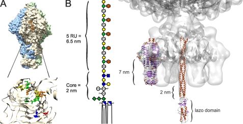
Initial attachment of bacteriophage P22 to the Salmonella host cell is known to be mediated by interactions between lipopolysaccharide (LPS) and the phage tailspike proteins (TSP), but the events that subsequently lead to DNA injection into the bacterium are unknown. We used the binding of a fluorescent dye and DNA accessibility to DNase and restriction enzymes to analyze DNA ejection from phage particles in vitro. Ejection was specifically triggered by aggregates of purified Salmonella LPS but not by LPS with different O-antigen structure, by lipid A, phospholipids, or soluble O-antigen polysaccharide. This suggests that P22 does not use a secondary receptor at the bacterial outer membrane surface. Using phage particles reconstituted with purified mutant TSP in vitro, we found that the endorhamnosidase activity of TSP degrading the O-antigen polysaccharide was required prior to DNA ejection in vitro and DNA replication in vivo. If, however, LPS was pre-digested with soluble TSP, it was no longer able to trigger DNA ejection, even though it still contained five O-antigen oligosaccharide repeats. Together with known data on the structure of LPS and phage P22, our results suggest a molecular model. In this model, tailspikes position the phage particles on the outer membrane surface for DNA ejection. They force gp26, the central needle and plug protein of the phage tail machine, through the core oligosaccharide layer and into the hydrophobic portion of the outer membrane, leading to refolding of the gp26 lazo-domain, release of the plug, and ejection of DNA and pilot proteins.


Figures

FIGURE 1.
FIGURE 1.
In vitro DNA ejection from phage P22 particles. To follow DNA ejection at 37 °C, we added 3.7 × 10⁹ phage P22 particles to 5 μg/ml S. typhimurium LPS and a fluorescent DNA-binding dye (●). Addition of DNase reversed the fluorescence increase, indicating DNA became released from the phage (○). When 11 nm free TSP were added 10 min after the phage particles, less DNA became ejected (▴). Neither LPS from E. coli (▾), digested S. typhimurium LPS (■), nor its lipid A mixed with O-antigen polysaccharide (×) was able to trigger DNA release from phage P22. Standard deviations from three independent experiments are not more than 4% of total fluorescence for every experiment.
FIGURE 2.
FIGURE 2.
Agarose gel electrophoresis of phage P22 and its ejection products. A, EtBr stained 1% agarose gel electrophoresis with 1.75 × 10⁹ complete P22 phages (lane 1) or complete phage particles with 10 μg/ml DNase (lane 2), 1.75 × 10⁹ phage particles incubated with LPS over night (lane 3), and thereafter with 10 μg/ml DNase (lane 4). B, electrophoretic analysis of DNA released from intact phage particles upon incubation with LPS (lanes 1, 3, and 5) and of phage DNA purified from denatured particles (lanes 2, 4, and 6), either untreated (lanes 1 and 2) or digested with AvaI (lanes 3 and 4) or ClaI (lanes 5 and 6).
FIGURE 3.
FIGURE 3.
TSP endorhamnosidase mutations delay DNA ejection from phage P22. The fluorescence ejection assay was used to follow DNA release from reconstituted phage particles carrying different TSP variants. To 10 μg/ml S. typhimurium LPS and the fluorescent dye, 7.2 × 10⁹ reconstituted phage particles (P22tmutant) were added at 37 °C as follows: ●, P22tWT; ▴, P22tT307K; ▾, P22tD392N; ■, P22tD395N. ○, fluorescence decrease after addition of DNase I; formula image
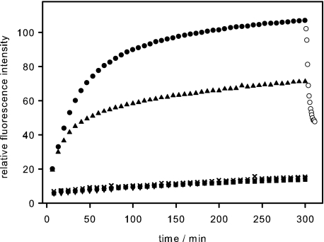
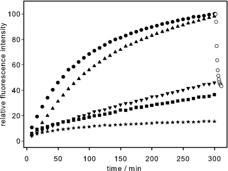
, P22tWT control without LPS. The standard deviation of fluorescence signals between repeated experiments was below 0.5%.
FIGURE 4.
FIGURE 4.
Binding and hydrolysis activity of TSP. A, fluorescence binding titration of S. typhimurium O-antigen polysaccharide at 10 °C. Tryptophan fluorescence of 0.11 μm TSPD392N was excited at 295 nm and quenching upon binding followed at 350 nm. B, kinetics TSP O-antigen polysaccharide cleavage. Samples were analyzed after the indicated times on a Superdex Peptide HR 10/30. C, 15% SDS-PAGE of LPS cleavage products. 15% silver-stained SDS-PAGE of purified LPS fraction (lane 1), LPS incubated with phage P22 (lane 2), and LPS digested with TSP and purified (lane 3).
FIGURE 5.
FIGURE 5.
Putative DNA release mechanism of phage P22 triggered by LPS. A, tailspike binding and activity site. Binding cleft in the trimeric protein of P22 TSP (9) is shown. Residues carrying the mutations at Thr-307 (green) and in the active site Asp-392 (red) and Asp-395 (blue) are indicated. B, cell attachment apparatus of phage P22. TSP and gp26 plug crystal structures were modeled into the cryo-EM structure of phage P22 (7, 32, 40, 53). Dimensions of the LPS digestion product containing five O-antigen repeats (38) match the distances in the model and suggest insertion of the gp26 lazo-domain into the hydrophobic part of the outer membrane, presumably resulting in its refolding. Sugar icons are according to Varki et al. (54), an orange symbol was added for the abequose.

References

    1. Prevelige P. E. (2006) in The Bacteriophages (Calendar R. ed) 2nd Ed., pp. 457–468, Oxford University Press, New York
    1. Israel J. V., Anderson T. F., Levine M. (1967) Proc. Natl. Acad. Sci. U.S.A. 57, 284–291 - PMC - PubMed
    1. Teschke C. M., Parent K. N. (2010) Virology 401, 119–130 - PMC - PubMed
    1. Seckler R. (1998) J. Struct. Biol. 122, 216–222 - PubMed
    1. Baxa U., Cooper A., Weintraub A., Pfeil W., Seckler R. (2001) Biochemistry 40, 5144–5150 - PubMed

Publication types

LinkOut - more resources