. 2011 Jan;39(1):347-58.
doi: 10.1093/nar/gkq749. Epub 2010 Sep 3.

Characterization of SMG-9, an essential component of the nonsense-mediated mRNA decay SMG1C complex

Affiliations

Characterization of SMG-9, an essential component of the nonsense-mediated mRNA decay SMG1C complex

Israel S Fernández et al. Nucleic Acids Res. 2011 Jan.

Abstract

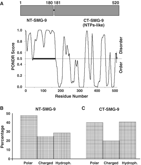
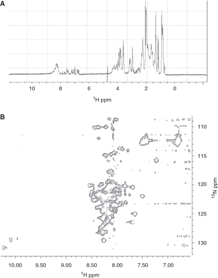
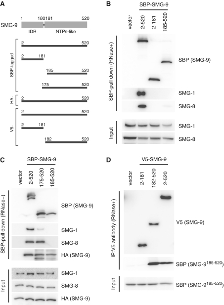
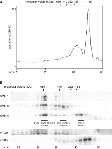
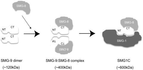
SMG-9 is part of a protein kinase complex, SMG1C, which consists of the SMG-1 kinase, SMG-8 and SMG-9. SMG1C mediated phosphorylation of Upf1 triggers nonsense-mediated mRNA decay (NMD), a eukaryotic surveillance pathway that detects and targets for degradation mRNAs harboring premature translation termination codons. Here, we have characterized SMG-9, showing that it comprises an N-terminal 180 residue intrinsically disordered region (IDR) followed by a well-folded C-terminal domain. Both domains are required for SMG-1 binding and the integrity of the SMG1C complex, whereas the C-terminus is sufficient to interact with SMG-8. In addition, we have found that SMG-9 assembles in vivo into SMG-9:SMG-9 and, most likely, SMG-8:SMG-9 complexes that are not constituents of SMG1C. SMG-9 self-association is driven by interactions between the C-terminal domains and surprisingly, some SMG-9 oligomers are completely devoid of SMG-1 and SMG-8. We propose that SMG-9 has biological functions beyond SMG1C, as part of distinct SMG-9-containing complexes. Some of these complexes may function as intermediates potentially regulating SMG1C assembly, tuning the activity of SMG-1 with the NMD machinery. The structural malleability of IDRs could facilitate the transit of SMG-9 through several macromolecular complexes.


Figures

Figure 1.
Figure 1.
Analysis of SMG-9 primary structure. (A) PONDR analysis of the SMG-9 amino acid sequence. A PONDR score >0.5 predicts those amino acids belonging to a disordered region. A stretch of disordered amino acids of more than 50 residues are usually considered a disordered domain. This analysis revealed that the sequence of SMG-9 could be divided in two regions: an N-terminal 180 residue intrinsically disordered region and a C-terminal folded domain, encoding a putative nucleotide-triphosphatase (NTPase)-like domain (12). Distribution of types of amino acid in NT-SMG-9 (B) and the C-terminal region of SMG-9 (C). NT-SMG-9 revealed a propensity towards polar and charged amino acids while the C-terminal domain was enriched in hydrophobic residues. Unstructured domains frequently exhibit a low content in hydrophobic amino acids and a bias towards charged and polar residues.
Figure 2.
Figure 2.
Expression and purification of NT-SMG-912–180 in E. coli. (A) SDS–PAGE of three different constructs assayed to produce a soluble fragment of NT-SMG-9. Left, N-terminal hexahistidine tag (pRHT vector); middle, periplasmic secretion vector (pRHO vector); and right, GST fusion protein (pGEX-6p-2 vector). The total cell extract right before induction (column T0), 5 h after induction with 1 mM IPTG (column T5) and the soluble fraction after sonication of the T5 sample (column S), are shown. For the periplasmic construct the supernatant (column SN) of the culture of T5 is shown, since the periplasmic space of E .coli is very leaky, over-expressed proteins can be easily localized in the supernatant of the centrifuged culture. Lastly, in those two constructs where some expression was detected, the elution from an in-batch incubation of the supernatant (periplasmic secretion construction) or the soluble fraction of T5 (GST-fusion construction) with HisTrap resin or GST–sepharose resin (column O), are shown. The pull-down protein in the GST-NT-SMG-9 construct is labelled. (B) SDS–PAGE of a purified NT-SMG-912–180 after the first GST-trap column before (column NC) and after a 4 h digestion (column C) with 3C protease. (C) Final preparation after applying the purification protocol described in the text.
Figure 3.
Figure 3.
Spectroscopic analyses of NT-SMG-912–180. (A) Far UV-circular dichroism spectrum of NT-SMG-912–180. (B) Fluorescence spectra of NT-SMG-912–180 in 50 mM phosphate buffer and 50 mM NaCl (black circles) and in the same buffer but in the presence of 6 M Guanidinium hydrochloride (white circles).
Figure 4.
Figure 4.
NMR spectroscopy analysis of NT-SMG-912–180. (A) 1H monodimensional spectrum of NT-SMG-912–180 showing the overlapping of signals in a narrow chemical shift in a range centered at 8.25 ppm. (B) 1H-15N HSQC spectrum of 15N-labelled NT-SMG-912–180 unambiguously identified this domain as inherently unstructured due to the absence of well dispersed cross peaks. The presence of well-resolved peaks and the absence of dispersion discarded any non-specific aggregation.
Figure 5.
Figure 5.
Effect of NT-SMG-9 and CT-SMG-9 truncation in SMG1C assembly. (A) Schematic structures of SMG-9 construct. (B) 293T cells were transfected with the SMG-9 plasmids shown above together with plasmid expressing the siRNA targeted to 3′-UTR of SMG-9. (C) 293T cells were transfected with the SBP-tagged-SMG-9 plasmids shown above together with the HA-tagged-SMG-9 plasmid. The cells were lysed and pull downed with the streptavidn sepharose in presence of RNaseA. Pull downed products or cell lysates (input) were then probed with the antibodies shown on the right. (D) 293T cells were transfected with the V5-tagged-SMG-9 plasmids shown above together with the SBP-tagged-CT-SMG-9185–520 plasmid. The cells were lysed and immunoprecipitated with anti-V5 antibodies in presence of RNaseA. Immunoprecipitated products or cell lysates (input) were then probed with the antibodies shown on the right. ‘Vector’ indicates an empty vector.
Figure 6.
Figure 6.
Several SMG-9-containing complexes can be isolated. (A and B) Size exclusion chromatography of SMG-1, SMG-8 and SMG-9 containing complexes. Fractions were run in SDS gels and the presence of either protein tested by western blot using anti-bodies specific to each component. As molecular weight markers, commercial markers (location of their elution peaks indicated in top line), mTOR (290 kDa and 0.7 MDa) and aPKCλ (78 kDa) were used.
Figure 7.
Figure 7.
Model for the assembly of SMG-9-containing complexes.

References

    1. Conti E, Izaurralde E. Nonsense-mediated mRNA decay: molecular insights and mechanistic variations across species. Curr. Opin. Cell Biol. 2005;17:316–325. - PubMed
    1. Isken O, Maquat LE. The multiple lives of NMD factors: balancing roles in gene and genome regulation. Nat. Rev. Genet. 2008;9:699–712. - PMC - PubMed
    1. Nicholson P, Yepiskoposyan H, Metze S, Zamudio Orozco R, Kleinschmidt N, Muhlemann O. Nonsense-mediated mRNA decay in human cells: mechanistic insights, functions beyond quality control and the double-life of NMD factors. Cell Mol Life Sci. 2009;67:677–700. - PMC - PubMed
    1. Brogna S, Wen J. Nonsense-mediated mRNA decay (NMD) mechanisms. Nat. Struct. Mol. Biol. 2009;16:107–113. - PubMed
    1. Rebbapragada I, Lykke-Andersen J. Execution of nonsense-mediated mRNA decay: what defines a substrate? Curr. Opin. Cell Biol. 2009;21:394–402. - PubMed

Publication types