. 2010 Oct 6;29(19):3344-57.
doi: 10.1038/emboj.2010.210. Epub 2010 Sep 3.

Proteomic and functional analysis of the mitotic Drosophila centrosome

Affiliations

Proteomic and functional analysis of the mitotic Drosophila centrosome

Hannah Müller et al. EMBO J. .

Abstract

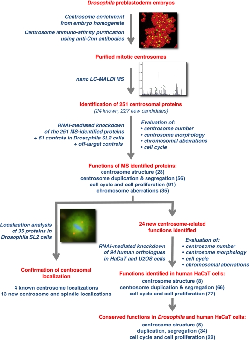
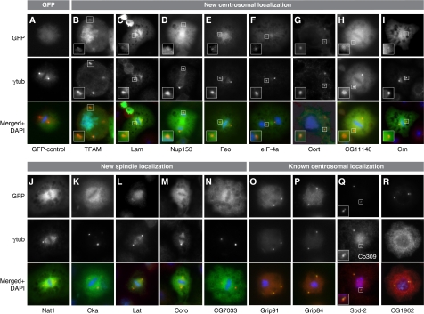
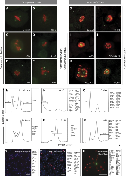
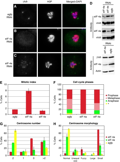
Regulation of centrosome structure, duplication and segregation is integrated into cellular pathways that control cell cycle progression and growth. As part of these pathways, numerous proteins with well-established non-centrosomal localization and function associate with the centrosome to fulfill regulatory functions. In turn, classical centrosomal components take up functional and structural roles as part of other cellular organelles and compartments. Thus, although a comprehensive inventory of centrosome components is missing, emerging evidence indicates that its molecular composition reflects the complexity of its functions. We analysed the Drosophila embryonic centrosomal proteome using immunoisolation in combination with mass spectrometry. The 251 identified components were functionally characterized by RNA interference. Among those, a core group of 11 proteins was critical for centrosome structure maintenance. Depletion of any of these proteins in Drosophila SL2 cells resulted in centrosome disintegration, revealing a molecular dependency of centrosome structure on components of the protein translation machinery, actin- and RNA-binding proteins. In total, we assigned novel centrosome-related functions to 24 proteins and confirmed 13 of these in human cells.


Conflict of interest statement

The authors declare that they have no conflict of interest.

Figures

Figure 1
Figure 1
Experimental approach and main findings of the proteomic and functional characterization of the early preblastoderm Drosophila centrosome. Drosophila preblastoderm embryo extract was used as starting material for the immunoisolation of mitotic centrosomes, followed by the identification of the centrosomal proteome components by mass spectrometry. The 251 identified proteins plus 61 controls were characterized by RNAi-mediated knockdown in Drosophila SL2 cells. Fifteen centrosomal, chromosomal and cell cycle features were analysed using immunofluorescence microscopy or FACS. Subsequently, localization analysis was performed (GFP-, TAP-tag expression and immunolocalization in SL2 cells) for the MS-identified proteins whose functional inhibition resulted in a ‘0' centrosome phenotype, for proteins with coiled-coil domains and for control proteins. Functional conservation of the identified proteins was confirmed in human HaCaT and U2OS cells. (Main experimental steps are shown in red colour, experimental procedures and main findings are shown in blue).
Figure 2
Figure 2
Confirmation of centrosomal or spindle localization of candidate MS-identified proteins and controls. Stable expression of GFP-fusion proteins in SL2 cells identifies new centrosomal and spindle localization of proteins whereas the GFP control shows uniform distribution (A). A centrosome associated localization was identified for TFAM (B), Lam (C), Nup153 (D) (transient expression), Feo (E), eIF-4a (F), Cort (G), CG11148 (H) and Crn (I). Not previously known was the spindle localization of the proteins Nat1 (J), Cka (K), Lat (L), Coro (M) and CG7033 (N). Positive controls confirm known centrosomal localization of Grip91 (O), Grip84 (P), Spd-2 (Q), CG1962 (R). The GFP-tag is shown in green (upper panels, AR), antibody staining against γ-tubulin (middle panels, AP, R) and Cp309 (Q) in red and superimposition of both images with DNA labelled by DAPI in blue (lower panels, AR). The inserts in BI and Q show a magnification of the area of the respective image marked with a white box.
Figure 3
Figure 3
Functional characterization of 251 MS-identified Drosophila centrosome candidate proteins plus 61 controls and 94 human orthologues identified centrosomal and cell cycle functions. (AL) Examples of the two phenotypic classes, aberrant centrosome structure (B, D, F, H, J, L) or centrosome duplication/segregation (C, E, I, K) revealed by RNAi-mediated knockdown in SL2 and HaCaT cells. The RNAi target protein is indicated within each panel. Anti-γ-tubulin (green) and anti-phospho-histone 3 (red) antibodies were used to label centrosomes and mitotic chromosomes, respectively. (MR) Examples of the cell cycle distribution profiles, determined by FACS analysis of dsRNA-treated SL2 cells. The RNAi target proteins whose depletion is inducing each phenotype are listed on the right of the corresponding cell cycle distribution profile. (M) Control (EGFP dsRNA-treated cells) cell cycle distribution, (N) Sub-G1, (O) G1/G0, (P) S-phase, (Q) G2/M, (R) more than G2 DNA content. (S, T) Representative fields of SL2 cells displaying low (S) or high (T) mitotic index following dsRNA treatment. The RNAi target proteins whose depletion is inducing each phenotype are listed on the right of the corresponding image. DAPI (blue) and anti-phospho-histone 3 antibodies (red) were used to label DNA and mitotic chromosomes, respectively. (U) Example of a cell showing an abnormal chromosome segregation phenotype. The RNAi target proteins, whose depletion results in an aberrant chromosome segregation phenotype are listed on the right of the image. Anti-γ-tubulin (green) and anti-phospho-histone 3 (red) antibodies were used to label centrosome and mitotic chromosomes, respectively. Scale bars represent 10 μm in (F, U), and 20 μm in (S, T). A complete list of all Drosophila and human proteins and the result of their functional analysis can be found in Supplementary Table S3.
Figure 4
Figure 4
The eukaryotic initiation factor 4a has a centrosome and cell cycle related function. As compared with the control (A), depletion of eIF-4a results in SL2 cells with small or no centrosomes as judged by staining with an γ-tubulin antibody (B, G, H), high mitotic index (E) and an accumulation of prophase cells (F), whereas eIF-4e knockdown led to cells with many centrosomes (C, G), normal mitotic index (E) and normal distribution of mitotic phases (F). Western blotting shows protein depletion by RNAi using anti-eIF-4a and eIF-4e antibodies and a stable protein level of γ-tubulin following eIF-4a knockdown. α-Tubulin and actin are used as loading controls (D). The distinct differences between the eIF-4a and eIF-4e RNAi phenotypes strongly suggest that the effect on the centrosome resulting from depletion of eIF-4a is most likely not a consequence of global inhibition of translation. The γ-tubulin reduction at the centrosome concomitant with an unaltered overall protein level in the cell indicates a mislocalization of γ-tubulin rather than disturbed translation (F, *could not be determined).

References

    1. Adams RR, Tavares AA, Salzberg A, Bellen HJ, Glover DM (1998) Pavarotti encodes a kinesin-like protein required to organize the central spindle and contractile ring for cytokinesis. Genes Dev 12: 1483–1494 - PMC - PubMed
    1. Andersen JS, Wilkinson CJ, Mayor T, Mortensen P, Nigg EA, Mann M (2003) Proteomic characterization of the human centrosome by protein correlation profiling. Nature 426: 570–574 - PubMed
    1. Badano JL, Teslovich TM, Katsanis N (2005) The centrosome in human genetic disease. Nat Rev 6: 194–205 - PubMed
    1. Bahmanyar S, Kaplan DD, Deluca JG, Giddings TH Jr, O'Toole ET, Winey M, Salmon ED, Casey PJ, Nelson WJ, Barth AI (2008) beta-Catenin is a Nek2 substrate involved in centrosome separation. Genes Dev 22: 91–105 - PMC - PubMed
    1. Barbosa V, Yamamoto RR, Henderson DS, Glover DM (2000) Mutation of a Drosophila gamma tubulin ring complex subunit encoded by discs degenerate-4 differentially disrupts centrosomal protein localization. Genes Dev 14: 3126–3139 - PMC - PubMed

Publication types

MeSH terms

Substances