. 2010 Oct;11(10):920-7.
doi: 10.1038/ni.1931. Epub 2010 Sep 5.

SLAM is a microbial sensor that regulates bacterial phagosome functions in macrophages

Affiliations

SLAM is a microbial sensor that regulates bacterial phagosome functions in macrophages

Scott B Berger et al. Nat Immunol. 2010 Oct.

Abstract

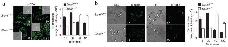
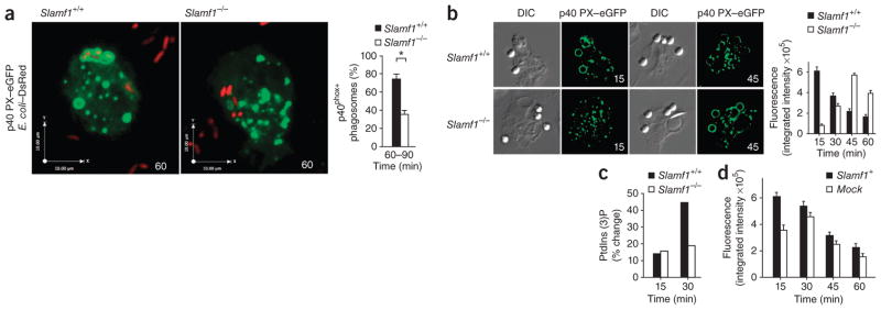
Phagocytosis is a pivotal process by which macrophages eliminate microorganisms after recognition by pathogen sensors. Here we unexpectedly found that the self ligand and cell surface receptor SLAM functioned not only as a costimulatory molecule but also as a microbial sensor that controlled the killing of gram-negative bacteria by macrophages. SLAM regulated activity of the NADPH oxidase NOX2 complex and phagolysosomal maturation after entering the phagosome, following interaction with the bacterial outer membrane proteins OmpC and OmpF. SLAM recruited a complex containing the intracellular class III phosphatidylinositol kinase Vps34, its regulatory protein kinase Vps15 and the autophagy-associated molecule beclin-1 to the phagosome, which was responsible for inducing the accumulation of phosphatidylinositol-3-phosphate, a regulator of both NOX2 function and phagosomal or endosomal fusion. Thus, SLAM connects the gram-negative bacterial phagosome to ubiquitous cellular machinery responsible for the control of bacterial killing.


Conflict of interest statement

COMPETING FINANCIAL INTERESTS

The authors declare no competing financial interests.

Figures

Figure 1
Figure 1
SLAM controls in vivo and in vitro killing of Gram-negative bacteria by mouse macrophages. (a,b) Bacteria in the spleens of Slamf1−/− Rag1−/− and Rag1−/− BALB/c mice (a) or Slamf1−/− and Slamf1+/+ BALB/c mice (b) 48 h after intraperitoneal injection of virulent S. typhimurium 14028s or attenuated S. typhimurium SseB. CFU, colony-forming units; ND, not detectable. Data are representative of four independent experiments (mean and s.d.). (ce) Killing of bacteria by peritoneal macrophages from Slamf1+/+ and Slamf1−/− BALB/c mice exposed to S. typhimurium SseB (c), E. coli F18 (d) or S. aureus (e), assessed by gentamycin assay. Data are representative of five independent experiments (mean and s.d.). (f) Uptake of bacteria by Slamf1+/+ (red solid lines) or Slamf1−/− (black solid lines) BALB/c peritoneal macrophages incubated at 37 °C with E. coli–eGFP or S. typhimurium–eGFP or by Slamf1+/+ macrophages incubated for 60 min at 4 °C with the bacteria (dotted lines). Right, mean fluorescence intensity (MFI). Data are representative of three independent experiments (mean and s.d.).
Figure 2
Figure 2
Defective NOX2 activity in primary macrophages derived from SLAM-deficient mice. (a) NOX2 activity in Slamf1+/+ and Slamf1−/− BALB/c peritoneal macrophages stimulated for 0–100 min with E. coli F18, S. aureus or PMA, assessed with lucigenin. Data are representative of five independent experiments (mean ± s.d.). (b) Phagosomal pH of Slamf1+/+, Slamf1−/− and gp91phox−/− B6 primary macrophages loaded for 0–200 min with pHrodo-coated E. coli or S. aureus, analyzed by flow cytometry. Data are representative of three independent experiments (mean ± s.d.). (c) NOX2 activity in primary macrophages in response to LPS, purified OmpC, peptidoglycan (PGN) or PMA, assessed with lucigenin. TLR4-KO, TLR4-deficient (strain del/Jtht; C3H). Data are representative of five independent experiments (mean ± s.d.).
Figure 3
Figure 3
Impaired phagolysosomal maturation in Slamf1−/− macrophages. (a) Fluorescence microscopy of E. coli–containing phagosomes in primary macrophages transfected with RFP-conjugated LAMP-1, showing colocalization with E. coli–eGFP or S. aureus–eGFP. Right, quantification of LAMP-1+ phagosomes in the microscopy at left. (b) Immunoblot analysis of LAMP-1 in phagosomes isolated by sucrose-gradient flotation from RAW264.7 macrophages after phagocytosis by Slamf1- or mock-transfected RAW264.7 macrophages of beads coated with E. coli outer membrane extract. WCL, whole-cell lysate. Right, quantification of LAMP-1 in the immunoblot at left. (c) Localization of MHC class II–eGFP (MHCII GFP) in phagosomes of primary macrophages from Slamf1+/+ or Slamf1−/− MHC class II–eGFP (B6) mice and 3-μm beads coated with E. coli outer membrane extract. (d) Entry of 3-μm beads coated with E. coli outer membrane extract into lysosomes loaded with Texas red–dextran. Right (c,d), quantification of fluorescence in the microscopy at left. Numbers in bottom right corners (a,c,d) indicate time (in min). Original magnification (ac), ×60. DIC, differential interference contrast. Data are representative of three combined experiments (a) or at least three independent experiments (bd) with at least 100 beads or 80 bacteria in each (error bars, s.d.).
Figure 4
Figure 4
Delay in early phagosomal maturation in Slamf1−/− macrophages. (a,b) Association of EEA-1 (a) or Rab5 (b) with phagosomes generated in primary macrophages by 3-μm beads coated with E. coli outer membrane extract; numbers in bottom corners indicate time (in min). α-, anti-. Original magnification, ×60. Right (a,b), quantification of fluorescence. Data are representative of at least three independent experiments (error bars, s.d.).
Figure 5
Figure 5
SLAM enters the E. coli–containing phagosome. (a) Colocalization of SLAM and bacteria in RAW264.7 macrophages transfected with SLAM-mCherry and allowed to phagocytose E. coli–eGFP or S. aureus–eGFP for 0–120 min (time (in min), bottom right corners at left). Original magnification, ×60. Right, quantification of fluorescence at left. Data are representative of two independent experiments with a minimum of 40 bacteria per time point (error bars, s.d.). (b) Immunoblot analysis of phagosome isolates from RAW264.7 macrophages transiently transfected with cDNA encoding Myc-tagged SLAM (Slamf1-Myc) or mock transfected and allowed to phagocytose beads coated with E. coli outer membrane extract for 60 or 120 min. kDa, kilodaltons. Data are representative of at least three independent experiments.
Figure 6
Figure 6
SLAM recognizes E. coli and S. typhimurium Sseb but not S. aureus. (a) Luciferase activity in Jurkat cells transfected with a fusion of SLAM and CD3ζ and a luciferase reporter (as described in Results), plus a renilla luciferase reporter, then exposed to heat-killed bacteria (top). (b) Luciferase activity in Jurkat cells transfected with the fusion in a (Slamf1-CD3ζ) or a fusion of SLAM ectodomain construct lacking the immunoglobulin V domain and CD3ζ (ΔIgV-Slamf1–CD3ζ), and luciferase reporters as in a, then exposed to heat-killed E. coli F18. (c) Luciferase activity in Jurkat cells transfected as in a, then left uninoculated (−) or inoculated with 10 × 108 E. coli F18 (+), followed by the addition of monoclonal antibody (mAb; amount, under graph) 9D1 to SLAM (α-SLAM) or rat immunoglobulin G2b isotype-matched control antibody (Rat IgG2b). (d) Luciferase activity in Jurkat cells transfected as in a, then exposed to medium alone (Med), wild-type E. coli (JM101), or JM101 E. coli mutants lacking both Omp C and Omp F (HN705) or lacking either OmpC (ΔOmpC) or OmpF (ΔOmpF). Data are representative of at least three independent experiments (error bars (a), s.d.).
Figure 7
Figure 7
PtdIns(3)P production in phagosomes of primary macrophages is controlled by SLAM. (a,b) Production of phagosomal PtdIns(3)P in E. coli–containing phagosomes of primary peritoneal macrophages transfected with reporter cDNA encoding an eGFP-tagged PX domain of p40phox (p40 PX–eGFP) and treated with DsRed-expressing E. coli (E. coli–DsRed; a) or beads coated with E. coli outer membrane extract (b). Numbers in bottom right corners indicate time (in min). Original magnification, ×60. Right, quantification of fluorescence in microscopy at left. (c) HPLC analysis of the production of PtdIns(3)P in primary macrophages labeled with 3H-tagged myoinositol and treated with beads coated with E. coli outer membrane extract. (d) PtdIns(3)P production in RAW264.7 cells stably expressing SLAM or a mock construct, transfected with eGFP-tagged p40 PX and treated with beads coated with E. coli outer membrane extract. Data are from three combined experiments (a) or are representative of at least three independent experiments (b,d) or two independent experiments (c) with at least 100 beads or bacteria per experiment (error bars, s.d.).
Figure 8
Figure 8
SLAM recruits the intracellular Vps34–Vps15–beclin-1 complex to the phagosome. (a,b) Immunoassay of 293 cells transfected with various combinations of full-length SLAM (a) or hemagglutinin-tagged (−HA) tailless SLAM (b), EAT-2A, V5-tagged (−V5) Vps34-Vps15, and beclin-1; proteins immunoprecipitated (IP) from lysates with monoclonal antibody to SLAM, as well as whole-cell lysates (WCL), were analyzed by immunoblot (IB) with anti-beclin-1, anti-Vps34, anti-V5, anti-hemagglutinin, anti-SLAM or anti-EAT2. (c) Microscopy of RAW264.7 cells transiently transfected with cDNA encoding GFP-tagged beclin-1 and SLAM-mCherry, then treated with beads coated with E. coli outer membrane extract (at a ratio of 10:1, beads/cells) and fixed after 60 min. Data are representative of six (a) or two (b,c) independent experiments.

References

    1. Stuart LM, Ezekowitz RA. Phagocytosis and comparative innate immunity: learning on the fly. Nat Rev Immunol. 2008;8:131–141. - PubMed
    1. Aderem A, Ulevitch RJ. Toll-like receptors in the induction of the innate immune response. Nature. 2000;406:782–787. - PubMed
    1. Calpe S, et al. The SLAM and SAP gene families control innate and adaptive immune responses. Adv Immunol. 2008;97:177–250. - PubMed
    1. Rethi B, et al. SLAM/SLAM interactions inhibit CD40-induced production of inflammatory cytokines in monocyte-derived dendritic cells. Blood. 2006;107:2821–2829. - PMC - PubMed
    1. Tatsuo H, Ono N, Tanaka K, Yanagi Y. SLAM (CDw150) is a cellular receptor for measles virus. Nature. 2000;406:893–897. - PubMed

Publication types

MeSH terms

Substances