. 2010 Nov;293(11):1971-83.
doi: 10.1002/ar.21242.

Ontogeny of the kidney and renal developmental markers in the rhesus monkey (Macaca mulatta)

Affiliations

Ontogeny of the kidney and renal developmental markers in the rhesus monkey (Macaca mulatta)

Cynthia A Batchelder et al. Anat Rec (Hoboken). 2010 Nov.

Abstract

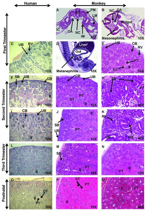
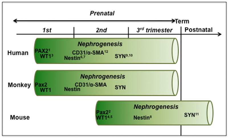
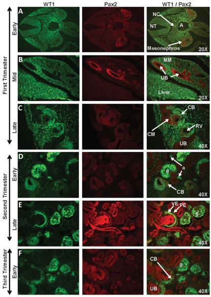
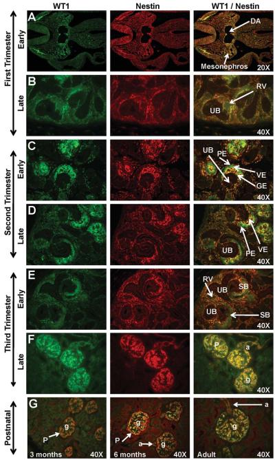
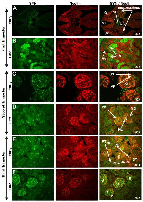
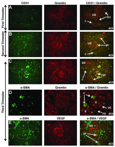
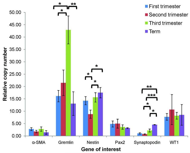
Nonhuman primates share many developmental similarities with humans, thus they provide an important preclinical model for understanding the ontogeny of biomarkers of kidney development and assessing new cell-based therapies to treat human disease. To identify morphological and developmental changes in protein and RNA expression patterns during nephrogenesis, immunohistochemistry and quantitative real-time PCR were used to assess temporal and spatial expression of WT1, Pax2, Nestin, Synaptopodin, alpha-smooth muscle actin (α-SMA), CD31, vascular endothelial growth factor (VEGF), and Gremlin. Pax2 was expressed in the condensed mesenchyme surrounding the ureteric bud and in the early renal vesicle. WT1 and Nestin were diffusely expressed in the metanephric mesenchyme, and expression increased as the Pax2-positive condensed mesenchyme differentiated. The inner cleft of the tail of the S-shaped body contained the podocyte progenitors (visceral epithelium) that were shown to express Pax2, Nestin, and WT1 in the early second trimester. With maturation of the kidney, Pax2 expression diminished in these structures, but was retained in cells of the parietal epithelium, and as WT1 expression was upregulated. Mature podocytes expressing WT1, Nestin, and Synaptopodin were observed from the mid-third trimester through adulthood. The developing glomerulus was positive for α-SMA (vascular smooth muscle) and Gremlin (mesangial cells), CD31 (glomerular endothelium), and VEGF (endothelium), and showed loss of expression of these markers as glomerular maturation was completed. These data form the basis for understanding nephrogenesis in the rhesus monkey and will be useful in translational studies that focus on embryonic stem and other progenitor cell populations for renal tissue engineering and repair.


Figures

Figure 1
Figure 1. Hematoxylin and Eosin (H&E) staining of sequential stages of kidney development
Sections of first trimester rhesus monkey embryos highlight development of the intermediate mesoderm (IM) (Fig. 1A; early first trimester transverse section), mesonephros (Fig. 1B; mid-first trimester transverse section), and definitive kidney or metanephros (Fig. 1D, sagittal section, and 1E; late first trimester). These developmental findings for nephrogenic structures were similar when compared to human fetal kidneys (8 weeks) (Fig. 1C) (courtesy of Dr. Douglas Matsell; collected in accordance with the Human Ethics Guidelines of the University of British Columbia). Second trimester images illustrate active nephrogenesis in the outer cortex in both human (Fig. 1F, I; 18 and 26 weeks, respectively; courtesy of D. Matsell) and monkey (Fig. 1G-H; early second trimester and Fig. 1J-K; mid-second trimester). Third trimester fetal kidney sections demonstrated nephrogenesis is nearing completion with restriction of the nephrogenic zone in human (Fig. 1L; 36 weeks; courtesy of D. Matsell) and monkey (Fig. 1M-N; mid-third trimester). Postnatal human (Fig. 1O; 5 years; courtesy of D. Matsell) and monkey kidneys (Fig. 1P-Q; 6 months) are shown. Abbreviations: CB, C-shaped body; DA, dorsal aorta; DT, distal tubule; IC, intraembryonic coelom; g, glomerulus; LM, lateral mesoderm; PM, paraxial mesoderm; NC, notochord; NT, neural tube; PT, proximal tubule; RV, renal vesicle; SB, S-shaped body; UB, ureteric bud.
Figure 2
Figure 2. Time course of nephrogenesis and expression of renal developmental markers in human, monkey, and mouse
Nephrogenesis begins in human and nonhuman primates in the late first trimester and continues throughout the mid-third trimester (term 165±10 days). In contrast, mouse nephrogenesis begins in mid-gestation and concludes postnatally (Saxen, 1987) (term 20 days). (1Tellier et al., 2000; 2Dressler et al., 1990; 3Charles et al., 1997; 4Armstrong et al., 1992; 5Rackley et al., 1993; 6Bertelli et al., 2007; 7Wagner et al., 2006; 8Chen et al., 2006, 9Nagata et al., 1998; 10Barisoni et al., 2000; 11Mundel et al., 1991; 12Naruse et al., 2000).
Figure 3
Figure 3. WT1 and Pax2 staining of sequential stages of kidney development in rhesus monkeys
Pax2 expression was noted from the early first trimester through the late third trimester in the mesonephros (Fig. 3A), ureteric bud (UB) (Fig. 3B), condensed mesenchyme (CM) (Fig. 3C-H), and renal vesicle (RV) (Fig. 3C) of the nephrogenic zone. Isolated collecting duct (CD) cells (Fig. 3I) continued to express Pax2 postnatally; this marker was also noted in widely scattered pockets of cells under the cortical capsule (Fig. 3I-J; 3-6 months postnatal WT1/Pax2 composite images). WT1 was expressed in the genital ridge between the aorta (A) and the mesonephros (Fig. 3A) and in the metanephric mesenchyme (MM) (Fig. 3B) in the mid-first trimester, followed by the C- and S-shaped bodies (CB and SB, respectively) (Fig. 3C-G), arterioles (a), and the visceral epithelium (VE) of the glomerulus where it was co-expressed with Pax2. The parietal epithelial layer (PE) did not express WT1 but showed isolated Pax2-positive cells that were closely associated with the vascular or urinary pole of the glomerulus (g) (Fig. 3E-G). Images oriented with the cortex to the upper left (Fig. 3C-K). NT, neural tube; NC, notochord; PT, proximal tubule.
Figure 4
Figure 4. WT1 and Nestin expression in sequential stages of kidney development in rhesus monkeys
In the early first trimester, WT1 was localized between the dorsal aorta (DA) and the mesonephros (Fig. 4A) while Nestin was expressed in the mesonephros. Dim expression of these markers was noted in the mid to late-first trimester metanephric kidney (Fig. 4B). Neither marker was expressed in the ureteric bud (UB). Faint WT1 expression was noted as the renal vesicle (RV) differentiated in the tail of the comma or C-shaped body. In the second trimester (Fig. 4C-D) and early third trimester (Fig. 4F), WT1 was strongly expressed on the visceral epithelium (VE) of the glomerulus with Nestin expression noted surrounding the developing glomerular endothelium (GE). The parietal epithelium (PE) of Bowman’s capsule was negative for both markers. By the mid-third trimester and continuing postnatally (Fig. 4F-G), these markers were expressed in the podocytes (P) of the glomeruli (g) and the afferent arterioles (a). Images oriented with cortex to the upper left. SB, S-shaped body. Postnatal (Fig. 4G) images are presented as a composite of WT1/Nestin.
Figure 5
Figure 5. Synaptopodin (SYN) and Nestin expression in sequential stages of kidney development in rhesus monkeys
In the early first trimester, Synaptopodin expression was noted in the notochord (NC) and surrounding region between the dorsal aorta (DA) and the neural tube (NT) (Fig. 5A). Nestin was localized to the mesonephros and the neural crest (arrows). Synaptopodin expression was observed in the ureteric bud (UB) in the late first trimester (Fig. 5B). Early second trimester expression of these markers was found in the visceral epithelial layer (VE) of mature glomeruli (g) deep in the medulla (Fig. 5C). Both markers were absent in the parietal epithelium (PE) of Bowman’s capsule. Synaptopodin expression in the outer cortical region was noted in late second trimester (Fig. 5D) on developing podocytes. This staining pattern intensified throughout the third trimester (Fig. 5E-F) where Synaptopodin and Nestin were co-expressed on podocytes (P). Images oriented with cortex to upper left. DT, distal tubule; MD, macula densa; RV, renal vesicle.
Figure 6
Figure 6. Markers of glomerular tuft development including CD31, Gremlin, alpha-smooth muscle actin (α-SMA), and vascular endothelial growth factor (VEGF) in sequential stages of nephrogenesis in rhesus monkeys
Dim CD31 immunoreactivity was observed on capillaries (c) in the metanephric blastema (Fig. 6A) while Gremlin staining was observed on the luminal surface of the ureteric bud (UB). Gremlin staining was stronger on the developing glomerular endothelium (ge) where it was co-expressed with CD31 in the second (Fig. 6B) and early third trimesters (Fig. 6C). By the mid-third trimester, only dim Gremlin expression was observed in a pattern consistent with endothelial or mesangial cells, and on the luminal surface of α-SMA-positive vasculature (Fig. 6D). Bright α-SMA expression was noted in vascular smooth muscle of arcuate arteries (not shown) and afferent arterioles (a). In the mid-second trimester, dim VEGF staining was noted followed by bright VEGF expression in third trimester glomerular endothelium (Fig. 6E). Images oriented with the cortex to upper left. PE, parietal epithelium; RV, renal vesicle; SB, S-shaped body; VE, visceral epithelium.
Figure 7
Figure 7. Quantitative RT-PCR analysis of expression patterns of α-SMA, Gremlin, Nestin, Pax2, Synaptopodin, and WT1 across gestation
RNA expression was quantified according to the comparative CT method with EF1-α as the housekeeping gene and relative changes in expression determined by comparison to adult mRNA taken as 100% (see text); *p<0.05, **p<0.01, ***p<0.001. An N≥3 was included for each age group.

References

    1. Agrawal R, Tran U, Wessely O. Expression and functional analysis of miRNAs in kidney development. Dev Biol. 2008;319:605. doi:10.1016/j.ydbio.2008.05.447.
    1. Agrawal R, Tran U, Wessely O. The miR-30 miRNA family regulates Xenopus pronephros development and targets the transcription factor Xlim1/Lhx1. Development. 2009;136:3927–3936. - PMC - PubMed
    1. Appel D, Kershaw DB, Smeets B, Yan G, Fuss A, Frye B, Elger M, Kriz W, Floege J, Moeller MJ. Recruitment of podocytes from glomerular parietal epithelial cells. J Am Soc Nephrol. 2009;20:333–343. - PMC - PubMed
    1. Armstrong JF, Pritchard-Jones K, Bickmore WA, Hastie ND, Bard JBL. The expression of the Wilms’ tumour gene, WT1, in the developing mammalian embryo. Mech Dev. 1992;40:85–97. - PubMed
    1. Batchelder CA, Lee CCI, Matsell DG, Yoder MC, Tarantal AF. Renal ontogeny in the rhesus monkey (Macaca mulatta) and directed differentiation of human embryonic stem cells towards kidney precursors. Differentiation. 2009;78:45–56. - PMC - PubMed

Publication types

MeSH terms