. 2010 Nov;162(2):315-24.
doi: 10.1111/j.1365-2249.2010.04243.x. Epub 2010 Sep 1.

The inhibitory effects of intravenous administration of rabbit immunoglobulin G on airway inflammation are dependent upon Fcγ receptor IIb on CD11c(+) dendritic cells in a murine model

Affiliations

The inhibitory effects of intravenous administration of rabbit immunoglobulin G on airway inflammation are dependent upon Fcγ receptor IIb on CD11c(+) dendritic cells in a murine model

M Yamamoto et al. Clin Exp Immunol. 2010 Nov.

Abstract

Immunoglobulins (Igs) play important immunomodulatory effects on allergic asthma. Among these, IgG has been reported to regulate allergic inflammation in previous studies about immunotherapy and intravenous immunoglobulin therapy. In this study, to examine the immunomodulatory mechanisms of IgG and FcRs we evaluated the effects of intravenous (i.v.) rabbit IgG administration (IVIgG) on allergic airway inflammation and lung antigen-presenting cells (APCs) in a murine model of ovalbumin (OVA) sensitization and challenge. In OVA-challenged mice, IVIgG attenuated airway eosinophilia, airway hyperresponsiveness and goblet cell hyperplasia and also inhibited the local T helper type (Th) 2 cytokine levels. Additionally, IVIgG attenuated the proliferation of OVA-specific CD4(+) T cells transplanted into OVA-challenged mice. Ex vivo co-culture with OVA-specific CD4(+) cells and lung CD11c(+) APCs from mice with IVIgG revealed the attenuated transcription level of Th2 cytokines, suggesting an inhibitory effect of IVIgG on CD11c(+) APCs to induce Th2 response. Next, to analyse the effects on Fcγ receptor IIb and dendritic cells (DCs), asthmatic features in Fcγ receptor IIb-deficient mice were analysed. IVIgG failed to attenuate airway eosinophilia, airway inflammation and goblet cell hyperplasia. However, the lacking effects of IVIgG on airway eosinophilia in Fcγ receptor IIb deficiency were restored by i.v. transplantation of wild-type bone marrow-derived CD11c(+) DCs. These results demonstrate that IVIgG attenuates asthmatic features and the function of lung CD11c(+) DCs via Fcγ receptor IIb in allergic airway inflammation. Targeting Fc portions of IgG and Fcγ receptor IIb on CD11c(+) DCs in allergic asthma is a promising therapeutic strategy.


Figures

Fig. 1
Fig. 1
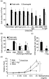
Effects of intravenous (i.v.) immunoglobulin G (IVIgG) on airway inflammation and hyperresponsiveness. (a) Ovalbumin (OVA)-sensitized mice received i.v. administration of rabbit IgG (IgG, 0·1–1000 µg), 1000 µg of IgM, F(ab′)2 or mouse IgG (m-IgG) 1 day before OVA challenge (OVA/OVA) and total cells and eosinophils in bronchoalveolar fluid (BALF) were quantified. (b) Schedules of rabbit IgG administration were compared. The number of total cells and eosinophils in BALF were measured in mice administered with rabbit IgG (1000 µg) before and after OVA challenge. (c) The level of plasma OVA-specific IgE in OVA-challenged mice was compared. (d) Airway hyperresponsiveness (AHR) was assessed using body plethysmography. At 24 h after the last challenge, increased enhanced pause (Penh) in response to inhaled methacholine were measured. The effects of IgG administration on AHR to methacholine were expressed on the Y-axis as relative values of Penh to baseline. *Significant differences (P < 0·05) versus naive mice. †Significant differences (P < 0·05), versus phosphate-buffered saline (PBS).
Fig. 2
Fig. 2
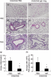
Effects of intravenous (i.v.) immunoglobulin G (IVIgG) on the development of airway inflammation and mucus production. Paraffin-embedded sections of lung of sensitized and challenged mice were prepared. Representative histological findings for lungs stained with haematoxylin and eosin (H&E) or periodic-acid Schiff (PAS) are shown. (a–c) Lung tissue from challenged mice [ovalbumin (OVA)/OVA + phosphate-buffered saline (PBS)]. (d–f) Lung tissue from mice administered with 1 mg of IgG (OVA/OVA + IgG). (a,d) Low-power field of the H&E-stained sample. (b,e) High-power field of the H&E-stained sample. (c,f) High-power fields of the PAS-stained sample, which shows mucus stained red by PAS. Scale bar equals 100 µm on low-power fields (a,d) and 50 µm on high-power fields (b,c,e,f). (g) Scores for peribronchial and perivascular inflammation are shown. (h) The number of airways free of mucus-producing cells or containing > 50% PAS-positive-cells are shown. *Significant differences (P < 0·05) versus PBS.
Fig. 3
Fig. 3
Effects of intravenous (i.v.) immunoglobulin G (IVIgG) on T helper type 2 (Th2) cytokines in bronchoalveolar fluid (BALF). Th1/Th2 cytokines in BALF were measured by enzyme-linked immunosorbent assay (ELISA). Interleukin (IL)-4 (a), IL-5 (b), IL-13 (c) and interferon (IFN)-γ (d) levels of ovalbumin (OVA)-sensitized and challenged mice are shown. *Significant differences (P < 0·05) versus naive mice. †Significant differences (P < 0·05) versus phosphate-buffered saline (PBS).
Fig. 4
Fig. 4
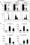
Effects of intravenous (i.v.) immunoglobulin G (IVIgG) on antigen presentation and T helper type 2 (Th2) differentiation. (a) Flow cytometric analysis of antigen presentation to ovalbumin (OVA)-specific CD4+ T cells in vivo is shown. Carboxyfluorescein succinimidyl ester (CFSE)-labelled CD4+ OVA-specific OTII lymphocytes (5 × 10⁶ cells) were transferred into mice challenged with OVA and administered with rabbit IgG or phosphate-buffered saline (PBS). At 24 h after the last challenge, the proliferation of CD4+ T cells in thoracic lymph nodes (TLNs) was analysed. The histogram shows the reduction of CFSE intensity, which indicates cell division and proliferation of the transferred CFSE+ CD4+ cells. One representative experiment of three with similar results is presented. In the inset boxes the percentages of divided (left box) or undivided cells (right box) among CFSE+ CD4+ cells subsets in representative data are shown. (a) The mRNA levels of Th2 cytokines [interleukin (IL)-4, IL-5, IL-13] and Th1 cytokine [interferon (IFN)-γ] in co-culture of OTII CD4+ T cells and lung CD11c+ cells with OVA323–339 peptide are shown. CD4+ cells (2·5 × 105 cells) isolated from spleens of OTII mice using CD4-microbeads and magnetic-activated cell sorting (MACS) system were stimulated with the OVA peptide (5 µg/ml) and lung CD11c+ cells (2·5 × 104 cells) from IgG/PBS-administered mice. After 6 h, mRNA levels of cytokines were evaluated by real-time polymerase chain reaction (PCR) *Significant differences (P < 0·05) versus naive mice. †Significant differences (P < 0·05) versus PBS.
Fig. 5
Fig. 5
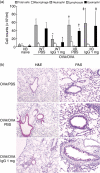
Effects of intravenous (i.v.) immunoglobulin G (IVIgG) on airway inflammation in FcγRIIb-deficient mice. (a) The cellular composition of bronchoalveolar fluid (BALF) in wild-type (WT) and FcγRIIb-deficient mice (IIB) are shown. Sensitized mice received IgG/ phosphate-buffered saline (PBS)-administration and challenged with ovalbumin (OVA). *Significant differences (P < 0·05) versus WT OVA/OVA + PBS, †versus IIB naive, ‡versus WT OVA/OVA + 1 mg IgG. (b) Representative histological findings of haematoxylin and eosin (H&E) or periodic-acid Schiff (PAS)-stained lung sections from FcγRIIb-deficient mice challenged with OVA are shown. Scale bar equals 50 µm.
Fig. 6
Fig. 6
Restoration of intravenous (i.v.) immunoglobulin G (IVIgG) effects on FcγRIIb-deficient mice by adoptive transfer of bone marrow-derived CD11c+ dendritic cells (BMDCs). The total cells and eosinophils in bronchoalveolar fluid (BALF) from FcγRIIb-deficient mice are shown. Sensitized and IgG or phosphate-buffered saline (PBS)-administered mice were transferred BMDCs derived from wild-type mice (WT) or FcγRIIb-deficient mice (IIB) before OVA challenge. *Significant differences (P < 0·05) versus OVA/OVA + BMDC (IIB) + IgG.

Similar articles

Cited by

References

    1. Wills-Karp M. Immunologic basis of antigen-induced airway hyperresponsiveness. Annu Rev Immunol. 1999;17:255–81. - PubMed
    1. Djukanovic R, Wilson SJ, Kraft M, et al. Effects of treatment with anti-immunoglobulin E antibody omalizumab on airway inflammation in allergic asthma. Am J Respir Crit Care Med. 2004;170:583–93. - PubMed
    1. Muller U, Akdis CA, Fricker M, et al. Successful immunotherapy with T-cell epitope peptides of bee venom phospholipase A2 induces specific T-cell anergy in patients allergic to bee venom. J Allergy Clin Immunol. 1998;101:747–54. - PubMed
    1. Haque S, Boyce N, Thien FC, O'Hehir RE, Douglass J. Role of intravenous immunoglobulin in severe steroid-dependent asthma. Intern Med J. 2003;33:341–4. - PubMed
    1. Salmun LM, Barlan I, Wolf HM, et al. Effect of intravenous immunoglobulin on steroid consumption in patients with severe asthma: a double-blind, placebo-controlled, randomized trial. J Allergy Clin Immunol. 1999;103:810–15. - PubMed

Publication types

MeSH terms