. 2010 Sep 6:10:18.
doi: 10.1186/1472-6793-10-18.

Increased expression and local accumulation of the prion protein, Alzheimer Aβ peptides, superoxide dismutase 1, and nitric oxide synthases 1 & 2 in muscle in a rabbit model of diabetes

Affiliations

Increased expression and local accumulation of the prion protein, Alzheimer Aβ peptides, superoxide dismutase 1, and nitric oxide synthases 1 & 2 in muscle in a rabbit model of diabetes

Claudine L Bitel et al. BMC Physiol. .

Abstract

Background: Muscle disease associated with different etiologies has been shown to produce localized accumulations of amyloid and oxidative stress-related proteins that are more commonly associated with neurodegeneration in the brain. In this study we examined changes in muscle tissue in a classic model of diabetes and hyperglycemia in rabbits to determine if similar dysregulation of Alzheimer Aβ peptides, the prion protein (PrP), and superoxide dismutase 1 (SOD1), as well as nitric oxide synthases is produced in muscle in diabetic animals. This wild-type rabbit model includes systemic physiological expression of human-like Alzheimer precursor proteins and Aβ peptides that are considered key in Alzheimer protein studies.

Results: Diabetes was produced in rabbits by injection of the toxic glucose analogue alloxan, which selectively enters pancreatic beta cells and irreversibly decreases insulin production, similar to streptozotocin. Quadriceps muscle from rabbits 16 wks after onset of diabetes and hyperglycemia were analyzed with biochemical and in situ methods. Immunoblots of whole muscle protein samples demonstrated increased PrP, SOD1, as well as neuronal and inducible Nitric oxide synthases (NOS1 and NOS2) in diabetic muscle. In contrast, we detected little change in Alzheimer Aβ precursor protein expression, or BACE1 and Presenilin 1 levels. However, Aβ peptides measured by ELISA increased several fold in diabetic muscle, suggesting a key role for Aβ cleavage in muscle similar to Alzheimer neurodegeneration in this diabetes model. Histological changes in diabetic muscle included localized accumulations of PrP, Aβ, NOS1 and 2, and SOD1, and evidence of increased central nuclei and cell infiltration.

Conclusions: The present study provides evidence that several classic amyloid and oxidative stress-related disease proteins coordinately increase in overall expression and form localized accumulations in diabetic muscle. The present study highlights the capacity of this wild-type animal model to produce an array of hallmark pathological features that have also been described in other muscle diseases.


Figures
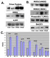
Figure 1
Figure 1
Increased expression of the Prion Protein (PrP), Superoxide dismutase 1 (SOD1), and Nitric oxide synthases 1 and 2, but not the Alzheimer Aβ precursor protein (AβPP), Presenilin 1 or BACE1 occurs in total muscle protein from diabetic rabbits. Immunoblot detection of proteins in diabetic and control muscle. A: PrP (MW ~30kDa, Epitomics), SOD1 (MW ~16kDa, Calbiochem), AβPP (MW ~110 kDa, Sigma), B: NOS1 (MW ~160 kDa, BD Biosciences), NOS2 (MW ~130 kDa, BD Biosciences), Presenilin 1 (MW ~52 kDa, Santa Cruz), BACE1 (MW ~55kDa, Santa Cruz) in total protein samples of quadriceps muscle from normal control (Con) and diabetic (Diab) rabbits. The doublet protein bands for PrP in diabetic and normal control muscle in Panel A is consistent with glycosylated forms, C: Densitometry measurements of protein bands shown on immunoblots above. Bar graphs show averages of the two band intensities with standard deviations. (NC: normal controls; D: diabetic).
Figure 2
Figure 2
ELISA assays measuring increased levels of Alzheimer Aβ peptides in diabetic muscle. Levels of Alzheimer Aβ peptides ending at amino acid 40 (x-40) or 42 (x-42) were determined in total muscle proteins from three diabetic and three normal control rabbits. ~7.5-fold increases were measured for Aβ40 and Aβ42 peptides in diabetic muscle compared to normal control muscle (P < 0.0005 and P < 0.0025, respectively). Graphs show pg/ml of Aβ detected in muscle samples determined using standard peptide controls (Covance). Graphs show mean values of 3 diabetic and 3 control animals with standard deviations.
Figure 3
Figure 3
Histological features generally characteristic of muscle degeneration linked with a variety of conditions are produced in quadriceps muscle from diabetic, hyperglycemic rabbits. Hematoxylin and eosin stained muscle sections from diabetic (A-F, H) and control (G, I) rabbits demonstrating central nuclei (A, H) and showing evidence of cell infiltration (A, C, F), not seen in normal control muscle. Muscle fiber cross-sections are ~40-50 μm in diameter. Images are representative of 3 diabetic and 3 control animals.
Figure 4
Figure 4
PrP, Aβ peptides, SOD1, NOS1 and NOS2 protein deposits accumulate in muscle fibers in diabetic rabbits. Above: Immunohistochemical peroxidase detection of A: Aβ peptides (mAb 6E10 Covance), B, C: PrP (rabbit mAb, Epitomics), D: NOS1 (mAb, BD Biosciences), E: NOS2 (mAb, BD Biosciences), F: Superoxide dismutase 1 (Calbiochem), G: No primary antibody, normal control, H: No primary antibody, diabetic muscle. Below: Immunofluorescence detection of proteins in diabetic muscle fibers. A: Aβ (mAb 4G8, Covance), B: PrP (mAb, Epitomics), C: overlay Aβ & PrP staining, D: Brightfield micrograph of degenerating muscle fiber showing vacuoles around the perimeter, E: Fluorescence photograph of anti-SOD1 staining in the same section as D, F: no primary antibody. Muscle fiber cross-sections are ~40-50 μm in diameter. Images are representative of 3 diabetic and 3 control animals.

References

    1. Barcelo A, Rajpathak S. Incidence and prevalence of diabetes mellitus in the Americas. Rev Panam Salud Publica. 2001;10(5):300–308. - PubMed
    1. Idiculla J, Shirazi N, Opacka-Juffry J, Ganapathi. Diabetic amyotrophy: a brief review. Natl Med J India. 2004;17(4):200–202. - PubMed
    1. Sun Z LL, Liu N, Liu Y. Muscular response and adaptation to diabetes mellitus. Front Biosci. 2008;13:4765–4794. doi: 10.2741/3038. - DOI - PubMed
    1. Yildirim Donmez FFF. Muscle compromise in diabetes. Acta Radiol. 2008;49:673–679. doi: 10.1080/02841850802105269. - DOI - PubMed
    1. Chen X GO, Geiger JD. Rabbits fed cholesterol-enriched diets exhibit pathological features of inclusion body myositis. Am J Physiol Regul Integr Comp Physiol. 2008;294:R829–835. - PubMed