

Insulin-induced hypoglycemic peripheral motor neuropathy in spontaneously diabetic WBN/Kob rats
- PMID: 20819377
- PMCID: PMC2930326
Insulin-induced hypoglycemic peripheral motor neuropathy in spontaneously diabetic WBN/Kob rats
Abstract
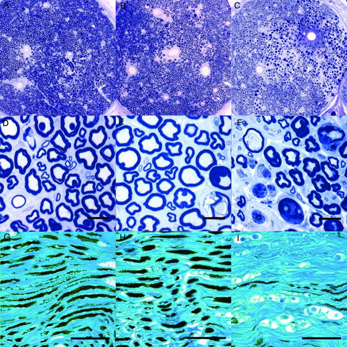
Intensive insulin therapy can lead to hypoglycemia, with patients sometimes developing hypoglycemic neuropathy. Spontaneously diabetic Wistar Bonn Kobori (WBN/Kob) rats develop diabetic peripheral motor neuropathy characterized by segmental demyelination and axonal degeneration. We examined the short-term effects of hypoglycemia on neuropathic changes in these rats. Spontaneous diabetic WBN/Kob rats received insulin implants for 40 d and were divided into 3 groups based on blood glucose levels: group N, normoglycemic to slightly hyperglycemic (150 to 250 mg/dL); group H, hypoglycemic to slightly hyperglycemic (50 to 200 mg/dL); and group D, nontreated spontaneously diabetic (350 to 420 mg/dL). Conduction velocity was measured in sciatic-tibial motor nerves; these nerves also underwent qualitative and quantitative histomorphologic analysis. Conduction velocity was not significantly different in N, D, and H groups. Morphologic analysis of the sciatic nerves of H rats showed severe changes, including axonal degeneration, myelin distention, and endoneurial fibrosis, that tended to occur in large, myelinated fibers. N and D rats showed relatively mild changes. The degree and distribution of degenerated nerve fibers in H rats were significantly higher than in N and D rats. These results suggest that hypoglycemia of less than 50 mg/dL induced severe peripheral neuropathy. Hypoglycemic lesions differed from the hyperglycemic lesions in diabetic WBN/Kob rats. This rat strain is an appropriate model for investigating the hypoglycemic peripheral neuropathy that can be associated with a diabetic condition.
Figures


References
-
- Grover-Johnson N, Spencer PS. 1981. Peripheral nerve abnormalities in aging rats. J Neuropathol Exp Neurol 40:155–165 - PubMed
-
- Ikegami H, Tabata H, Matsuzawa T, Suzuki H. 2000. The exacerbating effect of insulin-induced hypoglycemia on spontaneous peripheral neuropathy in aged B6C3F1 mice. J Toxicol Sci 25:137–142 - PubMed
-
- Jamali R, Mohseni S. 2006. Differential neuropathies in hyperglycemic and hypoglycemic diabetic rats. J Neuropathol Exp Neurol 65:1118–1125 - PubMed
-
- Low PA. 2005. Pathogenesis of diabetic neuropathy, p 839–851 : Kahn CR, Weir GC, King GL, Jakobson AM, Moses AC, Smith RJ. Joslin's diabetes mellitus. Boston (MA): Lipponcott Williams and Wilkins
Publication types
MeSH terms
Substances
LinkOut - more resources
Full Text Sources
Medical