. 2010 Aug 26;54(3):e39.
doi: 10.4081/ejh.2010.e39.

Histopathological and immunohistochemical study of the hepatic lesions experimentally induced by Entamoeba dispar

Affiliations

Histopathological and immunohistochemical study of the hepatic lesions experimentally induced by Entamoeba dispar

C A X Costa et al. Eur J Histochem. .

Abstract

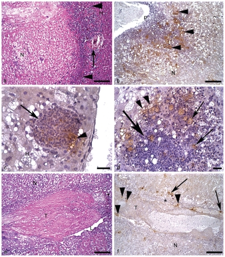
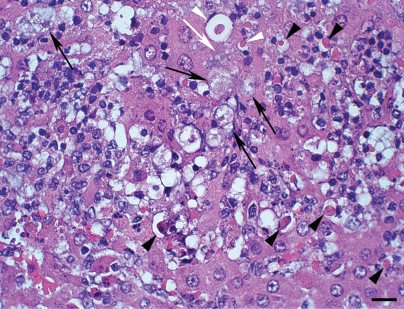
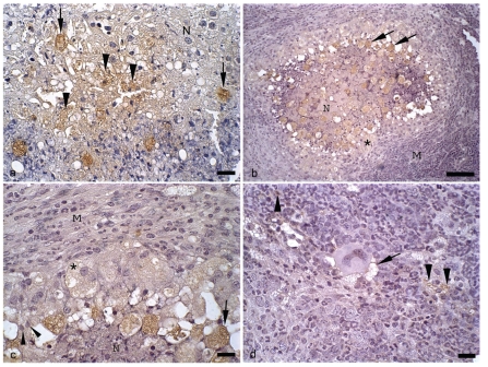
The sequence of hepatic necrotic-inflammatory events produced by Entamoeba dispar are originally described in this work. For the first time were described in details the experimental lesions produced by E. dispar, as well as the distribution of the trophozoites detected by the immunohistochemistry. Animals experimentally infected with E. dispar presented necrosis, thrombosis and chronic granulomatous inflammation. Immunoreactive products derived from trofozoites were observed close or associated with trophozoites, epithelioid cells, leucocytes and hepatocytes. Few are the articles on the literature about virulence of E. dispar, which is approximately 9 times more frequent than to E. histolytica. Variation in the virulence is, therefore expected and signalizing the need of the continuity of studies with E. dispar strains from different places in the world. Taking into account that E. dispar is a closely related species to E. histolytica, these studies could determine new elements involved with E. histolytica pathogenesis, helping us to understand better the disease.


Figures

Figure 1
Figure 1
Hamster liver inoculated with E. dispar. H&E and immunohistochemistry for E. dispar trophozoites, haematoxylin counterstained. (a) 1st day of infection. Necrosis (N); normal hepatic tissue (H); cell debris and scarce leucocytes (arrowheads); blood vessel partially obstructed by a thrombus (arrow). Bar 100 µm. H&E. (b) 2nd day of infection. Portal tract (P); numerous positive trophozoites for antigens of E. dispar (arrowheads); necrosis (N). Bar 100 µm. (c) 2nd day of infection. Granuloma rich in epithelioid cells (arrow) surrounded by degenerated hepatic tissue (H); note the positive reaction for E. dispar antigens inside the granuloma (arrowhead). Bar 20 µm. (d) 2nd day of infection. Granuloma constituted by epithelioid cells, macrophages and scarce lymphocytes (big arrow); stained trophozoites inside and close to the granuloma (arrows); positive immunohistochemistry reaction for E. dispar antigens inside vacuolated trophozoites (arrowheads). Bar 20 µm. (e) 2nd day of infection. Thrombus (T) almost totally occluding a branch of the portal vein; vascular lumen (L); necrosis of hepatic tissue (N). Bar 100 µm. H&E. (f) 2nd day of infection. Vascular wall (*); thrombus (T); stained trophozoites inside the thrombus (arrowheads) and close to the vascular wall (arrows); necrosis of hepatic tissue (N). Bar 100 µm.
Figure 2
Figure 2
Hamster liver inoculated with E. dispar (3rd day of infection) H&E. Hepatocytes show round shape, condensation of chromatin and cell shrinkage (arrowheads); trophozoites (arrows); note the presence of a trophozoite (white arrow) in close contact with hepatocyte shrinkage (white arrowheads). Bar 20 µm.
Figure 3
Figure 3
Hamster liver inoculated with E. dispar. and immunohistochemistry for E. dispar trophozoites, haematoxylin counterstained. (a) 6th day of infection. Presence of trophozoites (arrows) in area of hepatic necrosis (N); positive immunohistochemistry reaction for E. dispar antigens inside necrotic hepatocytes (arrowheads). Bar 20 µ m. (b) 8th day of infection. Granuloma with area of central necrosis (N) and several stained trophozoites (arrows); well compacted macrophages and epithelioid cells (*); mantle of concentrically arranged macrophages, lymphocytes and neutrophils (M). Bar 100 µm. (c) Higher view of the anterior figure showing foam macrophages with positive cytoplasmatic labeling for E. dispar antigens (*); epithelioid cells (arrow heads); trophozoite (arrow); mantle of macrophages, lymphocytes and neutrophils (M); necrosis (N). Bar 20 µm. (d) 8th day of infection. Detail of a mantle of macrophages, lymphocytes and neutrophils; note the positive reaction for E. dispar antigens inside inflammatory cells (arrowheads) and close to a Langhans-type giant cell (arrow). Bar 20 µm.

References

    1. Stanley SL., Jr Amoebiasis. Lancet. 2003;361:1025–1034. - PubMed
    1. Brumpt E. Etude sommaire de I'Entamoeba dispar n.sp. amibe akkystes quadrinuclees, parasite de I'homme. Bull Acad Méd Paris. 1925;94:942–52.
    1. Diamond LS. Amebiasis: a problem solved: What now? Arch Med Res. 1992;23:157–16. - PubMed
    1. Diamond LS, Clarck CG. A redescription of Entamoeba histolytica Schaudinn,1903 (Emended Walker, 1911) separating it from Entamoeba dispar Brumpt, 1925. J Eukaryot Microbiol. 1993;40:340–4. - PubMed
    1. Dodson JM, Clark CG, Lockhart LA, Schroeder JW, Mann BJ. Comparison of adherence, cytotoxicity, and Gal/GalNac lectin gene struture in Entamoeba histolityca and Entamoeba dispar. Parasitol Int. 1997;46:225–235.

Publication types