. 2010 Sep 27;207(10):2175-86.
doi: 10.1084/jem.20100637. Epub 2010 Sep 6.

Upregulation of Tim-3 and PD-1 expression is associated with tumor antigen-specific CD8+ T cell dysfunction in melanoma patients

Affiliations

Upregulation of Tim-3 and PD-1 expression is associated with tumor antigen-specific CD8+ T cell dysfunction in melanoma patients

Julien Fourcade et al. J Exp Med. .

Abstract

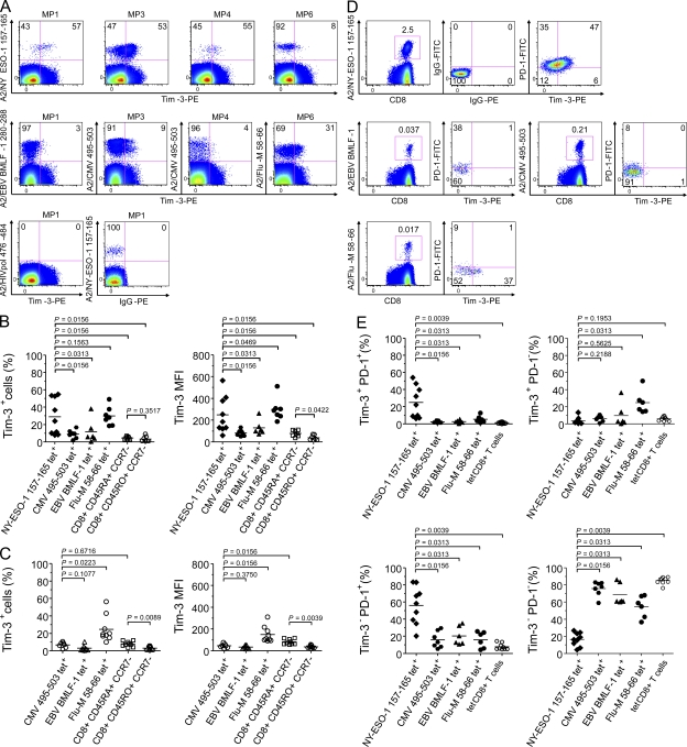
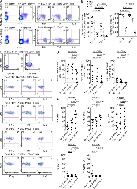
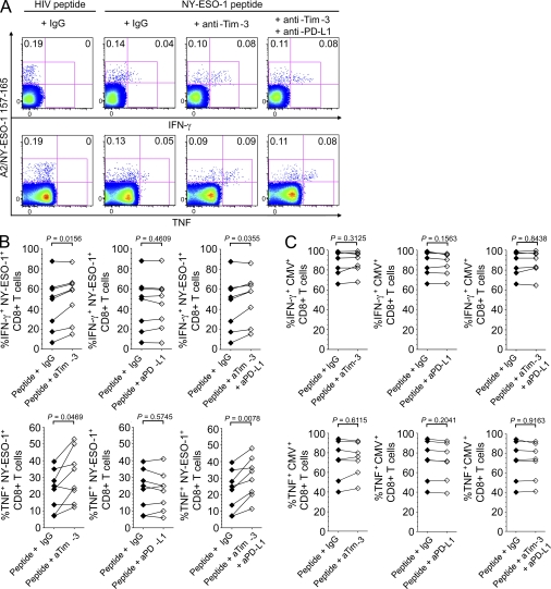
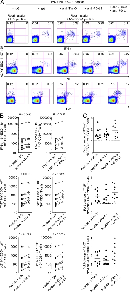
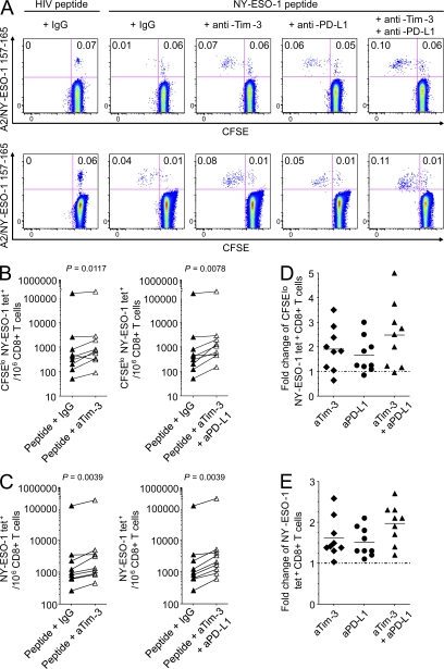
The paradoxical coexistence of spontaneous tumor antigen-specific immune responses with progressive disease in cancer patients furthers the need to dissect the molecular pathways involved in tumor-induced T cell dysfunction. In patients with advanced melanoma, we have previously shown that the cancer-germline antigen NY-ESO-1 stimulates spontaneous NY-ESO-1-specific CD8(+) T cells that up-regulate PD-1 expression. We also observed that PD-1 regulates NY-ESO-1-specific CD8(+) T cell expansion upon chronic antigen stimulation. In the present study, we show that a fraction of PD-1(+) NY-ESO-1-specific CD8(+) T cells in patients with advanced melanoma up-regulates Tim-3 expression and that Tim-3(+)PD-1(+) NY-ESO-1-specific CD8(+) T cells are more dysfunctional than Tim-3(-)PD-1(+) and Tim-3(-)PD-1(-) NY-ESO-1-specific CD8(+) T cells, producing less IFN-γ, TNF, and IL-2. Tim-3-Tim-3L blockade enhanced cytokine production by NY-ESO-1-specific CD8(+) T cells upon short ex vivo stimulation with cognate peptide, thus enhancing their functional capacity. In addition, Tim-3-Tim-3L blockade enhanced cytokine production and proliferation of NY-ESO-1-specific CD8(+) T cells upon prolonged antigen stimulation and acted in synergy with PD-1-PD-L1 blockade. Collectively, our findings support the use of Tim-3-Tim-3L blockade together with PD-1-PD-L1 blockade to reverse tumor-induced T cell exhaustion/dysfunction in patients with advanced melanoma.


Figures

Figure 1.
Figure 1.
Tim-3 is up-regulated and coexpressed with PD-1 on NY-ESO-1–specific CD8+ T cells. (A) Representative dot plots from melanoma patients (MP) showing ex vivo Tim-3 expression on A2/NY-ESO-1 157–165, A2/EBV BMLF-1 280–288, A2/CMV 495–503, and A2/Flu-M 58–66 tet+ CD8+ T cells. As shown for one melanoma patient (MP1), CD8+ T cells stained with A2/HIVpol 476–484 tetramers or PE-labeled IgG control Ab were used to establish the threshold for identifying tet+ cells and Tim-3+ cells, respectively. (B and C) Pooled data showing the percentage (%) and MFI of Tim-3 expression on NY-ESO-1–, CMV-, EBV-, and Flu-specific CD8+ T cells, as well as total effector (CD45RA+ CCR7) and effector/memory (CD45RO+ CCR7) CD8+ T cells from nine melanoma patients (B) and nine healthy donors (C). (D) Dot plots from one representative melanoma patient showing ex vivo Tim-3 and PD-1 expression on A2/NY-ESO-1 157–165, A2/EBV BMLF-1 280–288, A2/CMV 495–503 and A2/Flu-M 58–66 tet+ CD8+ T cells. As control, A2/NY-ESO-1 157–165 tet+ CD8+ T cells stained with PE-labeled and FITC-labeled IgG control antibodies are shown. (E) Pooled data showing the distribution of NY-ESO-1–, CMV-, EBV-, and Flu-specific CD8+ T cells, as well as total tet CD8+ T cells according to Tim-3 and PD-1 expression. Horizontal bars depict the mean percentage or MFI of Tim-3 and/or PD-1 expression on tet+ CD8+ T cells. The p- values were calculated using the Wilcoxon signed rank test. Data shown are representative of at least three independent experiments.
Figure 2.
Figure 2.
Co-expression of Tim-3 and PD-1 by NY-ESO-1–specific CD8+ T cells defines a population of dysfunctional T cells that up-regulate activation markers. (A and B) Representative dot plots from one melanoma patient (A) and summary data for melanoma patients (n = 7; B) showing the percentage of cytokine-producing A2/NY-ESO-1 157–165 tet+ and A2/Flu-M 58–66 tet+ CD8+ T cells among Tim-3+ and Tim-3 fractions after short ex vivo stimulation (6 h) with cognate peptide or irrelevant peptide (HIV peptide). (C and D) Dot plots from one patient (C) and summary data for all melanoma patients (n = 7; D) showing the percentages of cytokine-producing A2/NY-ESO-1 157–165 tet+ CD8+ T cells according to Tim-3 and PD-1 expression. (E) Pooled data from melanoma patients (n = 7) showing expression of CD38, HLA-DR, CD57, CCR7, and CD45RA on A2/NY-ESO-1 157–165 tet+ CD8+ T cells according to Tim-3 and PD-1 expression. The p-values were calculated using the Wilcoxon signed rank test. Data shown are representative of at least three independent experiments.
Figure 3.
Figure 3.
Ex vivo blockade of the Tim-3–Tim-3L pathway enhances cytokine production by NY-ESO-1–specific CD8+ T cells. (A and B) Representative dot plots from one melanoma patient (A) and summary data for all melanoma patients (n = 8; B) showing the percentages of A2/NY-ESO-1 157–165 tet+ CD8+ T cells that produce IFN-γ and TNF among total NY-ESO-1–specific CD8+ T cells after short ex vivo stimulation with cognate peptide in the presence of blocking anti–Tim-3 and/or anti–PD-L1 mAbs or an isotype control antibody (IgG). (C) Summary data for melanoma patients (n = 6) showing the percentages of A2/CMV 495–503 tet+ CD8+ T cells that produce IFN-γ and TNF among total CMV-specific CD8+ T cells after short ex vivo stimulation with cognate peptide in the presence of blocking anti–Tim-3 and/or anti–PD-L1 mAbs or an isotype control antibody (IgG). The p-values were calculated using the Wilcoxon signed rank test. Data shown are representative of two independent experiments performed in duplicate.
Figure 4.
Figure 4.
Blockade of the Tim-3–Tim-3L pathway alone or in combination with PD-1–PD-L1 blockade with prolonged antigen stimulation increases the frequency of cytokine-producing NY-ESO-1–specific CD8+ T cells. (A and B) Representative flow cytometry analysis from one melanoma patient showing percentages of IFN-γ-, TNF-, and IL-2–producing A2/NY-ESO-1 157–165 tet+ CD8+ T cells among total CD8+ T cells (A) and pooled data from melanoma patients (n = 9) showing the variation in the frequencies of IFN-γ–, TNF-, and IL-2–producing NY-ESO-1 tet+ cells for 106 CD8+ T cells (B). PBMCs were incubated for 6 d with NY-ESO-1 157–165 peptide or with HIVpol 476–484 peptide and blocking anti–Tim-3 (aTim-3) and/or anti–PD-L1 (aPD-L1) mAbs or an isotype control antibody (IgG) before evaluating intracellular cytokine production of A2/NY-ESO-1 157–165 tet+ CD8+ T cells in response to cognate peptide. (C) Fold change of the frequency of IFN-γ–, TNF-, and IL-2–producing A2/NY-ESO-1 157–165 tet+ CD8+ T cells after 6-d IVS with cognate peptide and blocking anti–Tim-3 and/or anti–PD-L1 mAbs. The ratio of the frequency of cytokine-producing tet+ CD8+ T cells from melanoma patients (n = 9) in the presence of indicated antibody treatment and isotype control antibody is shown. The p-values were calculated using the Wilcoxon signed rank test. Data shown are representative of two independent experiments performed in duplicate.
Figure 5.
Figure 5.
Blockade of the Tim-3–Tim-3L pathway alone or in combination PD-1–PD-L1 blockade with prolonged antigen stimulation increases the frequency of proliferating and total NY-ESO-1–specific CD8+ T cells. Representative flow cytometry analysis from two melanoma patients showing percentages of CFSElo A2/NY-ESO-1 157–165 tet+ CD8+ T cells among total CD8+ T cells (A) and pooled data from melanoma patients (n = 9) showing the variation in the numbers of CFSElo (B) and total (C) A2/NY-ESO-1 157–165 tet+ cells for 106 CD8+ T cells. CFSE-labeled PBMCs were incubated for 6 d with NY-ESO-1 157–165 peptide or HIVpol 476–484 peptide and blocking anti–Tim-3 (aTim-3), and/or anti–PD-L1 (aPD-L1) mAbs or an isotype control antibody (IgG). (D and E) Fold change of the frequencies of CFSElo (D) and total (E) A2/NY-ESO-1 157–165 tet+ CD8+ T cells after 6-d IVS with cognate peptide and blocking anti–Tim-3 (aTim-3) and/or anti–PD-L1 (aPD-L1) mAbs (n = 9). The ratio of the percentages of CFSElo and total A2/NY-ESO-1 157–165 tet+ CD8+ T cells in the presence of indicated antibody treatment and isotype control antibody is shown. The p-values were calculated using the Wilcoxon signed rank test. Data shown are representative of two independent experiments performed in duplicate.

Similar articles

Cited by

References

    1. Barber D.L., Wherry E.J., Masopust D., Zhu B., Allison J.P., Sharpe A.H., Freeman G.J., Ahmed R. 2006. Restoring function in exhausted CD8 T cells during chronic viral infection. Nature. 439:682–687 10.1038/nature04444 - DOI - PubMed
    1. Blackburn S.D., Shin H., Haining W.N., Zou T., Workman C.J., Polley A., Betts M.R., Freeman G.J., Vignali D.A., Wherry E.J. 2009. Coregulation of CD8+ T cell exhaustion by multiple inhibitory receptors during chronic viral infection. Nat. Immunol. 10:29–37 10.1038/ni.1679 - DOI - PMC - PubMed
    1. Boon T., Coulie P.G., Van den Eynde B.J., van der Bruggen P. 2006. Human T cell responses against melanoma. Annu. Rev. Immunol. 24:175–208 10.1146/annurev.immunol.24.021605.090733 - DOI - PubMed
    1. Day C.L., Kaufmann D.E., Kiepiela P., Brown J.A., Moodley E.S., Reddy S., Mackey E.W., Miller J.D., Leslie A.J., DePierres C., et al. 2006. PD-1 expression on HIV-specific T cells is associated with T-cell exhaustion and disease progression. Nature. 443:350–354 10.1038/nature05115 - DOI - PubMed
    1. Fourcade J., Kudela P., Andrade Filho P.A., Janjic B., Land S.R., Sander C., Krieg A., Donnenberg A., Shen H., Kirkwood J.M., Zarour H.M. 2008. Immunization with analog peptide in combination with CpG and montanide expands tumor antigen-specific CD8+ T cells in melanoma patients. J. Immunother. 31:781–791 10.1097/CJI.0b013e318183af0b - DOI - PMC - PubMed

Publication types

MeSH terms