. 2010 Sep 27;207(10):2097-111.
doi: 10.1084/jem.20101563. Epub 2010 Sep 6.

Inflammatory dendritic cells--not basophils--are necessary and sufficient for induction of Th2 immunity to inhaled house dust mite allergen

Affiliations

Inflammatory dendritic cells--not basophils--are necessary and sufficient for induction of Th2 immunity to inhaled house dust mite allergen

Hamida Hammad et al. J Exp Med. .

Abstract

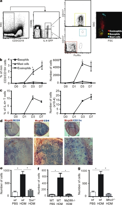
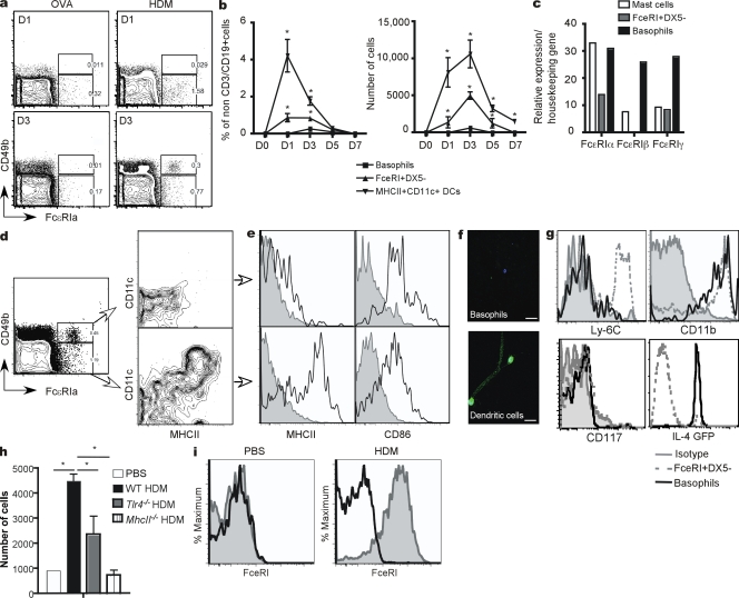
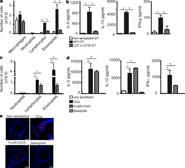
It is unclear how Th2 immunity is induced in response to allergens like house dust mite (HDM). Here, we show that HDM inhalation leads to the TLR4/MyD88-dependent recruitment of IL-4 competent basophils and eosinophils, and of inflammatory DCs to the draining mediastinal nodes. Depletion of basophils only partially reduced Th2 immunity, and depletion of eosinophils had no effect on the Th2 response. Basophils did not take up inhaled antigen, present it to T cells, or express antigen presentation machinery, whereas a population of FceRI(+) DCs readily did. Inflammatory DCs were necessary and sufficient for induction of Th2 immunity and features of asthma, whereas basophils were not required. We favor a model whereby DCs initiate and basophils amplify Th2 immunity to HDM allergen.


Figures

Figure 1.
Figure 1.
HDM exposure induces the recruitment of IL-4-competent basophils and eosinophils to the lung draining LNs. (a) Staining used to identify IL-4–competent cells in MLNs of HDM-administered animals. (b) Kinetics of recruitment of IL-4-EGFP+ basophils, mast cells, and eosinophils to the LNs after HDM administration. (c) IL-4-EGFP+ T cells at different time points after HDM administration. (d) Immunohistological staining of Mcpt8+ basophils (red) and CD4+ T cells, CD11c+ DCs, or B220+ cells (blue) in LNs at D3 after HDM administration. (e) Number of basophils recruited to the LNs of WT or Tlr4−/− mice 3 d after the administration of PBS or HDM. (f) Number of basophils recruited to the LNs of WT or Myd88−/− mice 3 d after the administration of PBS or HDM. (g) Number of basophils recruited to the LNs of WT or MhcII−/− mice 3 d after the administration of PBS or HDM. Data are representative of at least four independent experiments from four to six mice/group. Error bars represent the SEM. *, P < 0.05.
Figure 2.
Figure 2.
Contribution of basophils and eosinophils to Th2 immunity to inhaled HDM allergen. (a) Percentage of basophils in lung draining LNs of HDM-administered animals 3 d after the intravenous injection of anti-CD200R3 antibodies, anti-FcεRI antibodies, or control isotype-matched antibodies. Dot plots (IL4GFP vs CD49b) of lung draining LN cells gated on non– and -B cells. (b) Percentage of IL-4-EGFP+ Th2 cells in lung draining LNs of HDM- or PBS-administered animals 3 d after the intravenous injection of anti-FcεRI antibodies, anti-CD200R3 antibodies, or control isotype-matched antibodies. Dot plots (IL4GFP vs. CD3) of lung draining LN cells gated on CD3+ and lymphocyte scatter characteristics. Number of inflammatory cells in the BAL (c), presence of Siglec F+ eosinophils in the lungs (d), and cytokine production by LN cells of animals sensitized and challenged with HDM, and treated with anti- FcεRI or isotype-matched antibodies (e). (f) Number of inflammatory cells in the BAL, (g) presence of Siglec F+ eosinophils in the lungs, and (h) cytokine production by LN cells of animals sensitized and challenged with HDM, and treated with anti-CD200R3 or isotype-matched antibodies. Number of inflammatory cells in the BAL (i), presence of Siglec F+ eosinophils in the lungs (j), and cytokine production by LN cells of WT or eosinophil-deficient animals sensitized and challenged with HDM (k). Data are representative of at least three independent experiments from six to eight mice/group. Error bars represent the SEM. *, P < 0.05. Bars, 100 μm.
Figure 3.
Figure 3.
HDM exposure induces the recruitment of FcεRI-expressing monocyte-derived dendritic cells to MLNs. (a) Recruitment of FcεRI+DX5+ basophils and FcεRI+DX5 cells to the lung draining LNs 1 and 3 d after adminstration of OVA or OVA+HDM. (b) Kinetics of recruitment of FcεRI+DX5+ basophils, FcεRI+DX5 cells, and MHCII+CD11c+ DCs to the lung draining LNs after adminstration of HDM. (c) RT-PCR analysis of FcεRIα, β, and γ chain expression in FcεRI+DX5+ basophils and FcεRI+DX5 cells sorted from LNs 3 d after HDM administration, and of peritoneal mast cells of a naive mouse. (d) MHCII and CD11c expression by FcεRI+DX5+ basophils and FcεRI+DX5 cells in the LNs 3 d after the administration of HDM. (e) MHCII and CD86 expression by FcεRI+DX5+ basophils and FcεRI+DX5 cells in the LNs 3 d after the administration of HDM. (f) Immunofluorescence analysis of MHCII expression (green) on FcεRI+DX5+ basophils and FcεRI+DX5 cells sorted from the LNs of HDM-administered animals. Bars, 25 μm. DAPI (blue) was used to counterstain nuclei. Bars, 100 μm. (g) Ly-6C, CD11b, CD117 (c-Kit) and IL-4GFP expression by FcεRI+DX5+ basophils and FcεRI+DX5 cells in the LNs 3 d after the administration of HDM. (h) Number of FcεRI+DX5 cells recruited to the LNs of HDM- or PBS-administered animals. (i) FcεRI expression on lung MHCII+CD11c+ DCs 3 d after the administration of PBS (Left) or HDM (right). Solid black line, isotype-matched control antibody. Data are representative of at least three independent experiments from four to six mice/group. Error bars represent the SEM. *, P < 0.05.
Figure 4.
Figure 4.
FcεRI+ DCs, but not basophils, present antigens to T cells after exposure to HDM and induce Th2 immune responses. (a) OVA-AF647 uptake by FcεRI+DX5+ basophils (top) and FcεRI+DX5 cells (bottom) in the LNs 3 d after the administration of OVA-AF647 alone or in combination with HDM. (b) OVA-specific naive CD4+ T cell proliferation induced by FcεRI+DX5+ basophils, FcεRI+DX5 cells, or MHCII+CD11c+ DCs sorted from the LNs of OVA+HDM-administered animals 1 or 3 d after antigen exposure. (c) Cytokine production by OVA-specific naive CD4+ T cell restimulated for 4 d with FcεRI+DX5+ basophils, FcεRI+DX5 cells, or MHCII+CD11c+ DCs sorted from the LNs of OVA+HDM-administered animals 3 d after antigen exposure. (d) RT-PCR analysis of MHCII-associated chaperone proteins in FcεRI+DX5+ basophils, FcεRI+DX5 cells, or MHCII+CD11c+ DCs sorted from the LNs 3 d after HDM administration. Data are representative of at least three independent experiments from four to six mice/group.
Figure 5.
Figure 5.
Dendritic cells are necessary and sufficient for Th2 responses to HDM allergen. (a) Number of inflammatory cells in the BAL and (b) cytokine production by LN cells of WT or CD11c-DTR animals sensitized and challenged with HDM and treated with DT on day 0. (c) Number of inflammatory cells in the BAL, and (d) Cytokine production by LN cells of animals injected on day 0 i.p with basophils, FcεRI+DX5 cells or MHCII+CD11c+ DCs sorted from the lung draining LNs 1 d after HDM administration. Mice were then challenged with HDM on days 7–11. (e) Immunofluorescence staining of Siglec F+ eosinophils (red) in the lungs. DAPI (blue) was used to counterstain nuclei. Data are representative of at least two independent experiments from four to six mice/group. Error bars represent the SEM. *, P < 0.05. Bars, 100 μm.
Figure 6.
Figure 6.
Induction of Th2 immunity is a feature of FcεRI+ inflammatory type DCs. (a) Number of inflammatory cells in the BAL of mice sensitized by an intratracheal administration of DCs generated from BM cultured in the presence of GM-CSF or Flt3-liter. (b) FcεRI expression on DC subsets generated from BM cultured in the presence of GM-CSF or Flt3-liter. (c) Staining of FcεRI, CD49b, MHCII, and CD11c on BM cells cultured for 10 d with 30 ng/ml IL-3. (d) FcεRI expression on sorted monocytes after 1 or 3 d of culture with GM-CSF or IL-3. Data are representative of at least four independent experiments from four to eight mice/group. Error bars represent the SEM. *, P < 0.05.

References

    1. Allenspach E.J., Lemos M.P., Porrett P.M., Turka L.A., Laufer T.M. 2008. Migratory and lymphoid-resident dendritic cells cooperate to efficiently prime naive CD4 T cells. Immunity. 29:795–806 10.1016/j.immuni.2008.08.013 - DOI - PMC - PubMed
    1. Banchereau J., Steinman R.M. 1998. Dendritic cells and the control of immunity. Nature. 392:245–252 10.1038/32588 - DOI - PubMed
    1. Barrett N.A., Austen K.F. 2009. Innate cells and T helper 2 cell immunity in airway inflammation. Immunity. 31:425–437 10.1016/j.immuni.2009.08.014 - DOI - PMC - PubMed
    1. Barrett N.A., Maekawa A., Rahman O.M., Austen K.F., Kanaoka Y. 2009. Dectin-2 recognition of house dust mite triggers cysteinyl leukotriene generation by dendritic cells. J. Immunol. 182:1119–1128 - PMC - PubMed
    1. Baumeister T., Rössner S., Pech G., de Bruijn M.F., Leenen P.J., Schuler G., Lutz M.B. 2003. Interleukin-3Ralpha+ myeloid dendritic cells and mast cells develop simultaneously from different bone marrow precursors in cultures with interleukin-3. J. Invest. Dermatol. 121:280–288 10.1046/j.1523-1747.2003.12380.x - DOI - PubMed