. 2010 Sep 6;190(5):853-66.
doi: 10.1083/jcb.201005117.

Functional dichotomy of ribosomal proteins during the synthesis of mammalian 40S ribosomal subunits

Affiliations

Functional dichotomy of ribosomal proteins during the synthesis of mammalian 40S ribosomal subunits

Marie-Françoise O'Donohue et al. J Cell Biol. .

Abstract

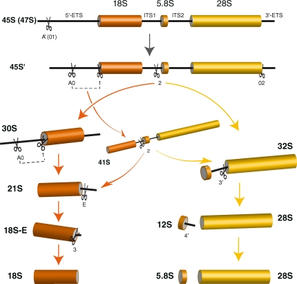
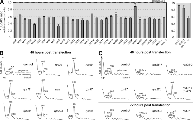
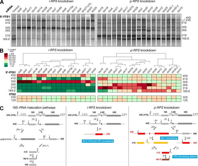
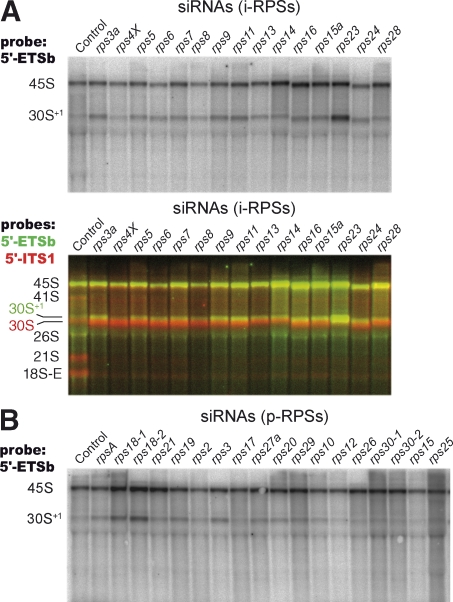
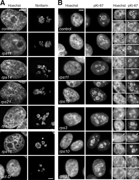
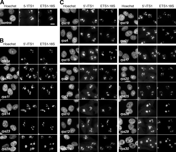
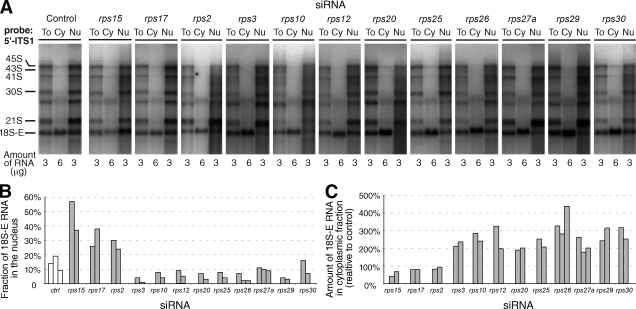
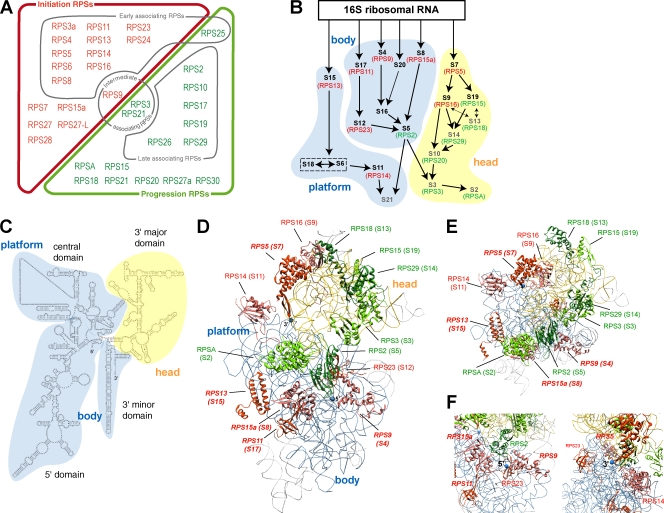
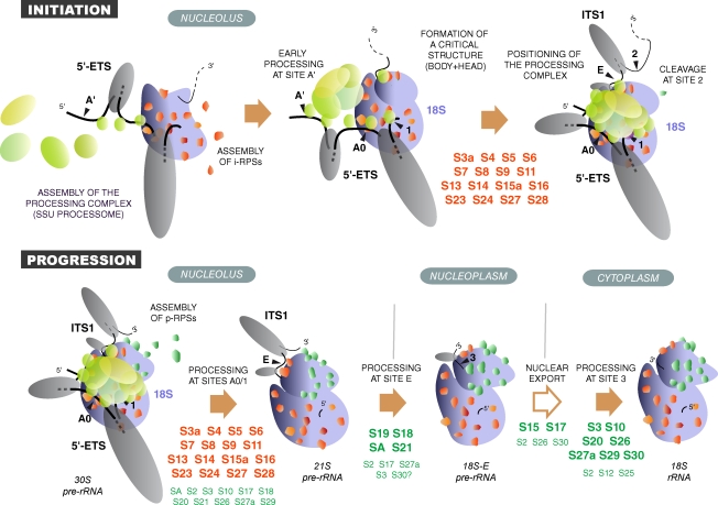
Our knowledge of the functions of metazoan ribosomal proteins in ribosome synthesis remains fragmentary. Using siRNAs, we show that knockdown of 31 of the 32 ribosomal proteins of the human 40S subunit (ribosomal protein of the small subunit [RPS]) strongly affects pre-ribosomal RNA (rRNA) processing, which often correlates with nucleolar chromatin disorganization. 16 RPSs are strictly required for initiating processing of the sequences flanking the 18S rRNA in the pre-rRNA except at the metazoan-specific early cleavage site. The remaining 16 proteins are necessary for progression of the nuclear and cytoplasmic maturation steps and for nuclear export. Distribution of these two subsets of RPSs in the 40S subunit structure argues for a tight dependence of pre-rRNA processing initiation on the folding of both the body and the head of the forming subunit. Interestingly, the functional dichotomy of RPS proteins reported in this study is correlated with the mutation frequency of RPS genes in Diamond-Blackfan anemia.


Figures

Figure 1.
Figure 1.
Pre-rRNA processing pathways in human cells. Two pathways coexist depending on the order of cleavage in the 5′-ETS (sites A0 and 1) and ITS1 (site 2). The major pathway in HeLa cells involves cleavage at site 2 before processing of ITS1, as shown by the higher abundance of 30S pre-rRNA relative to 41S pre-rRNA (see Fig. 3). The 18S-E pre-rRNA is converted to 18S rRNA in the cytoplasm. Nomenclature of the cleavage sites follows Hadjiolova et al. (1993) and Rouquette et al. (2005). The A’ early cleavage site is also designated 01.
Figure 2.
Figure 2.
Knockdown of each RPS protein affects 40S particle production. (A) Total RNAs were extracted from HeLa cells 48 h after transfection with siRNAs targeting RPS mRNAs. The 18S/28S ratio was calculated from Northern blot analysis to estimate the impact of each siRNA on 40S subunit production compared with control cells. Means of three to eight independent experiments with two different siRNAs ± SD are shown. (B) Cytoplasmic fractions were prepared in the presence of cycloheximide from cells transfected for 48 h with rps siRNAs or with a scrambled siRNA. Depletion of RPS proteins leads to the loss of free 40S particles and accumulation of free 60S particles, indicating a strong alteration in 40S subunit production. For each target protein, similar results were obtained with both siRNAs. (C) Knockdown of RPS25, RPS27, or RPS27L for 72 h, instead of 48 h, had no additional effect.
Figure 3.
Figure 3.
Analysis of pre-rRNA profiles upon individual knockdown of RPS proteins reveals their sequential role during 40S particle formation. Total RNAs from HeLa cells were extracted 48 h after transfection, resolved on a 1% agarose gel, and transferred to a nylon membrane, which was hybridized with various 32P-labeled probes (5′-ITS1, ITS2, 18S, or 28S). (A) A representative result with the 5′-ITS1 probe is shown for each RPS protein. The proteins fall into two categories: i-RPSs act in the early steps of processing (left), whereas p-RPSs impact a broad range of 18S rRNA maturation steps (right). (B) Hierarchical clustering of the pre-rRNA processing phenotypes after principal component analysis. Pre-rRNAs were quantified on Northern blots by phosphoimaging and normalized to the amount of 28S. The relative abundance of the precursors detected either with the 5′-ITS1 probe (top) or the ITS2 probe (bottom) relative to control cells is indicated by the color code (percentage of control level). Each column in the table corresponds to the mean of two to eight experiments and mixes the data obtained with two different siRNAs for most genes. siRNAs rps30-1 and rps30-2 yielded slightly different results but were very close in the clustering. (C) Alteration of the 18S rRNA maturation pathways in mammalian cells on knockdown of i-RPSs or p-RPSs. (left) Processing of the large 47S primary transcript can follow two alternative paths. (middle) Upon depletion of i-RPSs, all cleavages beyond that at site 2 were no longer taking place, leading to major accumulation of 30S pre-rRNAs. (right) When p-RPSs were knocked down, the 30S pre-rRNAs were at least partly processed, leading to 18S-E pre-rRNA production, and a wider range of phenotypes was observed: blocked or delayed processing at sites A0, 1, E, and 2 (accumulation of 30S, 21S, and 18S-E) and uncoupling of cleavage at sites A0 and 1 (accumulation of 26S and 45S). Red, strong accumulation; yellow, moderate accumulation; green, under accumulation.
Figure 4.
Figure 4.
Processing of the early cleavage site in the 5′-ETS of the primary transcript is still taking place upon depletion of RPS proteins. Pre-rRNAs uncleaved at site A’ were detected with probe 5′-ETSb, spanning nucleotides 301–324. This probe predominantly revealed the 45S pre-rRNA together with an extended form of the 30S pre-rRNA that we called 30S+1. (A) The 30S+1 species accumulated slightly on depletion of i-RPS proteins. However, the majority of the 30S RNA detected with probe 5′-ITS1 corresponds to a faster migrating band (overlay), indicating that it is processed at site A’. (B) Mild accumulation of 30S+1 RNA was observed on depletion of some p-RPSs only, especially RPS18.
Figure 5.
Figure 5.
Nucleolar morphology and chromatin condensation are modified by the depletion of specific RPSs. (A) Nucleolar morphology visualized with mAb 72B9. Fibrillarin is distributed in the dense fibrillar component, which outlines the fibrillar centers (small dots). Depletion of RPSs leads to changes in the number and shape of the nucleoli. Upon depletion of RPS16, nucleoli show partial segregation of the fibrillar components, similar to what is observed on RNA polymerase I inhibition for 4 h with 5 nM actinomycin D. (B) Knockdown of RPSs affected chromatin condensation and localization. Perinucleolar chromatin, visualized with antibodies to pKi-67, appeared as a rim around nucleoli in control HeLa cells (arrowheads). Upon knockdown of some proteins, like RPS5, RPS11, or RPS16, this rim was interrupted and disorganized (arrows show some examples). Depletion of other RPSs, like RPS3, RPS10, or RPS21, resulted in the redistribution of chromatin within nucleoli, as also seen by Hoechst staining of nucleoli, which was as intense as in the nucleoplasm. Bars, 5 µm.
Figure 6.
Figure 6.
Intracellular localization of the precursors to the 40S subunit in RPS-depleted cells. Pre-rRNAs of the 18S rRNA maturation pathway were detected by FISH with probes 5′-ITS1 (Cy3) and ETS1-18S (Cy5). (A–C) Control cells (A), representative examples of cells in which i-RPSs have been depleted (B), and cells in which p-RPSs have been knocked down (C). These FISH experiments were systematically performed in parallel with Northern blot analyses. Images were captured in identical conditions, and gray levels were scaled within the same lower and upper limits, except for RPS23-depleted cells for which the upper limit was twice that of the other images because of a much lower signal. The 5′-ITS1 labeling is displayed with a γ value of 1.5 to enhance the lowest gray levels. Bar, 10 µm.
Figure 7.
Figure 7.
Nucleocytoplasmic distribution of the 18S rRNA precursors in p-RPS–depleted cells. (A) At 48 h after transfection with siRNAs, cytoplasmic and nuclear RNAs were isolated and analyzed by Northern blotting. A representative experiment shows the patterns obtained after hybridization with the 5′-ITS1 probe for total extracts (To), the cytoplasmic fractions (Cy), and nuclear fractions (Nu). The amount of RNA loaded on the gel was 3 µg/well for total and nuclear extracts and 6 µg/well for cytoplasmic extracts. (B) Percentage of 18S-E pre–rRNA found in the nuclear fraction, as calculated from the balance of the whole fractionation procedure. The levels of 18S-E were measured with a phosphoimager. (C) Amount of 18S-E rRNA in the cytoplasmic fraction relative to control. The amount of 18S-E rRNA in the cytoplasmic fractions was normalized according to the amount of 28S rRNA (measured after hybridization with a specific probe on the phosphoimager), and siRNA-treated cells were compared with control cells. (B and C) The results of two or three independent experiments are shown for each RPS.
Figure 8.
Figure 8.
Distribution of i-RPSs and p-RPSs in the 40S ribosomal subunit. (A) Functional classification of RPSs according to their role in pre-rRNA processing. This classification is compared with the association order established in previous experiments for a large number of RPSs (see Discussion; Hadjiolov, 1985). RPSs shown to associate early belong to the i-RPS set (red), except for RPS25, whereas late associating proteins correspond to p-RPSs (green). (B) Position of i-RPSs and p-RPSs bacterial homologues in the assembly map of the 30S subunit (adapted from Holmes and Culver, 2004). Arrows between ribosomal proteins indicate their mutual dependency for association. Primary and secondary binding ribosomal proteins are shown in black and tertiary ribosomal proteins in gray. Human homologues are indicated by brackets (red, i-RPSs; green, p-RPSs). Correspondence of the ribosomal protein nomenclatures in Homo sapiens, S. cerevisiae, and Escherichia coli is provided in Table S2. (C) Secondary structure of the murine 18S rRNA. (D) Position of the i-RPS (in red or salmon red) and p-RPS (in dark and light green) proteins in the model of the canine mammalian ribosome (Protein Data Bank accession no. 2ZKQ) based on cryoelectron microscopy (Chandramouli et al., 2008). The 18S rRNA head domain is colored in yellow, whereas the body is in blue. Because localization of ribosomal proteins in eukaryotic models relies on the crystal structure of bacterial ribosomal subunits, only the 15 RPS proteins having a homologue in bacteria are positioned. The names of the corresponding bacterial proteins are indicated in parentheses. Proteins homologous to bacterial primary binders are in bold, italic characters. Pictures were produced using Chimera (Pettersen et al., 2004). (E) Top view of the head showing arrangement of p-RPSs. (F) Closer view of the 18S rRNA 5′ and 3′ ends (blue dots) shows that they are surrounded by i-RPSs.
Figure 9.
Figure 9.
Model of RPS protein activity in ribosome biogenesis. i-RPS proteins (red) assemble with the nascent pre-rRNA, most likely cotranscriptionally (Chooi and Leiby, 1981), together with early preribosomal factors (blue), in particular UTP proteins and sno-RNPs, like U3 and U17. i-RPS proteins participate in the folding of the 18S rRNA domain, whereas large secondary structures form in the transcribed spacers (Renalier et al., 1989; Michot and Bachellerie, 1991), potentially involving the binding of preribosomal factors. This allows formation of an assembly intermediate in which the processing machinery is correctly positioned, thus initiating cleavage of the 5′-ETS. p-RPSs (green) are mostly involved in downstream steps, although they may be necessary for coordination of processing at sites A0 and 1. The pre-40S particles are released from the nucleolus after cleavage at site E and are exported to the cytoplasm where the final processing step occurs. The RPS proteins required at each processing step are indicated.

Similar articles

Cited by

References

    1. Adilakshmi T., Bellur D.L., Woodson S.A. 2008. Concurrent nucleation of 16S folding and induced fit in 30S ribosome assembly. Nature. 455:1268–1272 10.1038/nature07298 - DOI - PMC - PubMed
    1. Auger-Buendia M.A., Longuet M., Tavitian A. 1979. Kinetic studies on ribosomal proteins assembly in preribosomal particles and ribosomal subunits of mammalian cells. Biochim. Biophys. Acta. 563:113–128 - PubMed
    1. Barkić M., Crnomarkovic S., Grabusić K., Bogetić I., Panić L., Tamarut S., Cokarić M., Jerić I., Vidak S., Volarević S. 2009. The p53 tumor suppressor causes congenital malformations in Rpl24-deficient mice and promotes their survival. Mol. Cell. Biol. 29:2489–2504 10.1128/MCB.01588-08 - DOI - PMC - PubMed
    1. Beltrame M., Tollervey D. 1995. Base pairing between U3 and the pre-ribosomal RNA is required for 18S rRNA synthesis. EMBO J. 14:4350–4356 - PMC - PubMed
    1. Borovjagin A.V., Gerbi S.A. 2001. Xenopus U3 snoRNA GAC-box A’ and box A sequences play distinct functional roles in rRNA processing. Mol. Cell. Biol. 21:6210–6221 10.1128/MCB.21.18.6210-6221.2001 - DOI - PMC - PubMed

Publication types

MeSH terms