. 2010 Nov 1;588(Pt 21):4089-101.
doi: 10.1113/jphysiol.2010.194738.

Cotransport of water by the Na+-K+-2Cl(-) cotransporter NKCC1 in mammalian epithelial cells

Affiliations

Cotransport of water by the Na+-K+-2Cl(-) cotransporter NKCC1 in mammalian epithelial cells

Steffen Hamann et al. J Physiol. .

Abstract

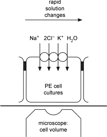
Water transport by the Na+-K+-2Cl(-) cotransporter (NKCC1) was studied in confluent cultures of pigmented epithelial (PE) cells from the ciliary body of the fetal human eye. Interdependence among water, Na+ and Cl(-) fluxes mediated by NKCC1 was inferred from changes in cell water volume, monitored by intracellular self-quenching of the fluorescent dye calcein. Isosmotic removal of external Cl(-) or Na+ caused a rapid efflux of water from the cells, which was inhibited by bumetanide (10 μm). When returned to the control solution there was a rapid water influx that required the simultaneous presence of external Na+ and Cl(-). The water influx could proceed uphill, against a transmembrane osmotic gradient, suggesting that energy contained in the ion fluxes can be transferred to the water flux. The influx of water induced by changes in external [Cl(-)] saturated in a sigmoidal fashion with a Km of 60 mm, while that induced by changes in external [Na+] followed first order kinetics with a Km of about 40 mm. These parameters are consistent with ion transport mediated by NKCC1. Our findings support a previous investigation, in which we showed water transport by NKCC1 to be a result of a balance between ionic and osmotic gradients. The coupling between salt and water transport in NKCC1 represents a novel aspect of cellular water homeostasis where cells can change their volume independently of the direction of an osmotic gradient across the membrane. This has relevance for both epithelial and symmetrical cells.


Figures

Figure 1
Figure 1. Basic experimental set-up used to measure water transport in cultured pigmented ciliary epithelial cells
The basolateral membranes of cultured pigmented epithelial (PE) cells from the ciliary body of the eye face upwards, and express isoform 1 of the Na+ −K+ −2Cl cotransporter (NKCC1). The water transport properties of the basolateral membranes are derived from the initial rates of change in cell water volume produced by rapid changes in ion concentrations of the bathing solution, at constant osmolarity, or by changing the external osmolarity (e.g. with mannitol). At the beginning of each experiment the cells are loaded with the fluorescent dye calcein; changes in cell volume are derived from self-quenching of this fluorophore monitored though the microscope's objective lens.
Figure 2
Figure 2. Removal of external Cl in isosmotic media produced bumetanide-sensitive shrinkage of pigmented ciliary epithelial cells
Isosmotic replacement of Cl with gluconate caused rapid cell shrinkage. Vt/Vo denotes relative cell water volume. Black bars above each trace indicate the time of exposure to the isosmotic Cl-free solution (0 Cl). Dashed vertical lines indicate onset of exposure to 0 Cl. On returning to the Cl-containing control solution cells recovered their initial volume. Right trace: the initial rate of cell shrinkage was decreased in the presence of 10 μm bumetanide.
Figure 5
Figure 5. Ion requirements and sensitivity to bumetanide and calyculin-A of net water fluxes across the basolateral membrane
Compiled data were obtained from experiments like those illustrated in Figs 2, 3 and 4. Net water fluxes were calculated from the initial rates of change in cell water volume and from measurements in cell height using eqn (2). The water efflux on exposure to the isosmotic Cl-free solution (Cl-removal) was reduced significantly (P < 0.001) by exposure to 10 μm bumetanide (Cl-removal (bum)). The net water influx measured on returning Cl to the bath (Cl-addition) was not significantly different from the net water efflux measured on removal of Cl, but was reduced significantly (P < 0.001) by bumetanide (Cl-addition (bum)). Similarly, the net water efflux induced on exposure to the isosmotic Na+-free solution (Na+-removal) was significantly (P < 0.001) reduced in the presence of bumetanide (Na+-removal (bum)). The water influx measured on returning Na+ to the bath (Na+-addition) was not significantly different from that obtained on removal of Na+; the water influx, however, was significantly (P < 0.001) reduced by bumetanide (Na+-addition (bum)). The largest net water fluxes observed were those elicited on isosmotic removal of external Cl in cells treated with 10 nm calyculin-A (Cl-removal (caly)), a phosphatase inhibitor that stimulates NKCC1. Calyculin-A caused a twofold increase that was statistically significant (P < 0.001).
Figure 3
Figure 3. Effect of calyculin-A on pigmented ciliary epithelial cell shrinkage produced by isosmotic removal of external Cl
Isosmotic replacement of Cl with gluconate caused rapid cell shrinkage (left trace). The initial rate of cell shrinkage increased significantly in cells treated with 10 nm calyculin-A, a phosphatase inhibitor known to stimulate NKCC1. Labels and symbols as in Fig. 2.
Figure 6
Figure 6. Net water influx as a function of external Cl and Na+ concentrations
A, pigmented ciliary epithelial cells were equilibrated with isosmotic Cl-free solution (0 Cl). Then, they were exposed to single pulses of isosmotic solutions each of which had a different Cl concentration (29.5, 59, 88.5, or 118 mm). At the end of each exposure the cells were returned to the Cl-free solution. Net water influx (formula image) for each pulse was estimated from the initial rates of cell swelling using eqn (2). Each data point represents the mean ±s.e.m. of 6 experiments. Data were fitted by a sigmoid curve with a Hill coefficient of about 2. B, cells were equilibrated with isosmotic Na+-free solution (0 Na+) and exposed to pulses of isosmotic solutions of increasing Na+ concentrations: 28.1, 56.2, 84.3, or 112.4 mm. At the end of each exposure the cells were returned to the Na+-free solution. Net water influx (formula image) for each pulse was estimated from the initial rates of cell swelling using eqn (2). Each data point represents the mean ±s.e.m. of 5 experiments. Data were fitted by a Michaelis–Menten equation with an apparent Km value of about 40 mm.
Figure 4
Figure 4. Isosmotic removal of external Na+ produced bumetanide-sensitive shrinkage of pigmented ciliary epithelial cells
Isosmotic replacement of Na+ with N-methyl-d-glucamine caused rapid cell shrinkage. Black bars above each trace indicate the time of exposure to the isosmotic Na+-free solution (0 Na+). Dashed vertical lines indicate onset of exposure to 0 Na+. On returning to the Na+-containing control solution, cells recovered their initial volume. Right trace: the initial rate of cell shrinkage was decreased in the presence of 10 μm bumetanide. Labels and symbols as in Fig. 2.
Figure 7
Figure 7. Water transport in pigmented ciliary epithelial cells requires the simultaneous presence of external Cl and Na+
A, left trace: isosmotic removal of external Cl (black bar, 0 Cl) caused an immediate, rapid cell shrinkage that reversed on returning to the Cl-containing bathing solution (vertical dashed line). Right trace: a second exposure to the 0 Cl solution produced a similar cell shrinkage to that observed in the left trace. While the cells were shrunken, they were exposed to isosmotic Na+-free solution (black bar, 0 Na+) containing the control concentration of Cl. In the absence of external Na+ there was no recovery from shrinkage (open arrow and dashed vertical line). Thus, on restoring the transmembrane Cl gradient in the absence of external Na+ there is no cell water volume recovery. On returning to the control isosmotic solution the cells recovered their initial water volume. B, isosmotic removal of external Na+ (black bar, 0 Na+) caused immediate, rapid cell shrinkage that reversed on returning to the control isosmotic solution (left vertical dashed line). While the cells were shrunken, they were exposed to isosmotic Cl-free solution (top black bar, 0 Cl) containing the control Na+ concentration. In the absence of external Cl there was no recovery from shrinkage even though the Na+ gradient had been restored (open arrow and dashed vertical line). On returning to the control isosmotic solution the cells recovered their initial water volume.
Figure 8
Figure 8. The Cl-dependent influx of water in pigmented ciliary epithelial cells can proceed against an osmotic gradient
Following equilibration in control solution, cells were exposed to isosmotic Cl-free solution, which resulted in cell shrinkage. On readmission of the Cl-containing control solution, cells recovered from shrinkage due to water influx (arrow 1 and dashed line). Addition of 50 mm mannitol to the control bathing solution (50 man), thereby rendering it about 17% hyperosmotic with respect to the control solution, resulted in osmotic shrinkage due to rapid cell water efflux (arrow 2 and vertical dashed line). On readmission of the control solution, cells recovered their initial water volume. However, when shrunken in isosmotic Cl-free solution, and then exposed to the Cl-containing control solution with 50 mm of mannitol added, cells increased their relative volume due to a rapid water influx (arrow 3 and dashed line). That is, water influx induced by concomitant Cl influx proceeded uphill, against the osmotic gradient imposed by 50 mm mannitol. On returning to isosmotic Cl-free solutions, cells went back to their initial (shrunken) volume, and on admission of the control solution, cells’ water volume recovered to its initial value.
Figure 9
Figure 9. Effect of bumetanide or changes in external ion composition on osmotic water permeability of the basolateral membrane of pigmented ciliary epithelial cells
A, addition of mannitol to the control bathing solution (indicated by the vertical broken line and the black bar), produced cell shrinkage. The water permeability (LP) was derived from the initial rate of cell shrinkage measured within the first 5–10 s of the response, applying eqn (2). In the example shown, 50 mosmol l−1 of mannitol was added to the control bathing solutions during the time indicated by the black bar. Vt/V0 denotes relative cell water volume. B, effect of bumetanide or changes in ion composition on Lp measured by applying hyperosmotic mannitol challenges (open bars) or NaCl (hatched bars). Bumetanide (10 μm) reduced Lp to half its control value (Control + bum) in mannitol. In tissues equilibrated with isosmotic Na+-free solution (0 Na+), Lp was reduced to about one-third of the control value. A similar reduction in Lp was measured in tissues equilibrated with isosmotic Cl-free solution (0 Cl). If the transmembrane osmotic gradients were changed by hyperosmotic NaCl (hatched bars), the Lp values were about half those obtained using hyperosmotic mannitol challenges (Control, hatched bar). Bumetanide had no effect on Lp values determined using hyperosmotic NaCl (Control + bum, hatched bar). C, diagram illustrating the two water pathways proposed to be present in the basolateral membrane of PE cells.
Figure 10
Figure 10. A molecular model of salt and water coupling in secretory epithelial cells
In this model, the primary coupling between salt and water transport takes place in the NKCC1 protein located in the serosal (basolateral) membrane of the secretory epithelial cell. The secondary active import of Na+, K+, Cl and H2O mediated by NKCC1 is energized by the Na+ gradient generated and maintained by the Na+/K+-ATPase. Na+ entering the cells via NKCC1 is pumped out by the Na+/K+-ATPase. K+ entering via NKCC1 leaks out through channels. Water and Cl proceed through the cell. Water leaves passively across the luminal (apical) membrane, e.g. via aquaporins (AQP), whereas Cl leave via channels (or transporters) in the apical membrane. Our data suggests that 570 water molecules and 2 Cl are cotransported per cycle via NKCC1. Na+ ions are secreted paracellularly via leaky junctions. For simplicity other cellular or paracellular pathways are not shown.

Comment in

References

    1. Agre P, Nielsen S, Ottersen OP. Towards a molecular understanding of water homeostasis in the brain. Neuroscience. 2004;12:849–850. - PubMed
    1. Alvarez-Leefmans FJ. Chloride transporters in presynaptic inhibition, pain and neurogenic inflammation. In: Alvarez-Leefmans FJ, Delpire E, editors. Physiology and Pathology of Chloride Transporters and Channels in the Nervous System: From Molecules to Diseases. San Diego, CA: Elsevier-Academic Press; 2009.
    1. Alvarez-Leefmans FJ, Altamirano J, Crowe WE. Use of ion-selective microelectrodes and fluorescent probes to measure cell volume. Methods Neurosci. 1995;27:361–391.
    1. Alvarez-Leefmans FJ, Gamino SM, Giraldez F, Nogueron I. Intracellular chloride regulation in amphibian dorsal root ganglion neurones studied with ion-selective microelectrodes. J Physiol. 1988;406:225–246. - PMC - PubMed
    1. Charron FM, Blanchard MG, Lapointe J-Y. Intracellular hypertonicity is responcible for water flux associated with Na+/glucose cotransport. Biophys J. 2006;90:3546–3554. - PMC - PubMed

Publication types

MeSH terms