

Human ocular carotenoid-binding proteins
- PMID: 20820671
- PMCID: PMC3938892
- DOI: 10.1039/c0pp00126k
Human ocular carotenoid-binding proteins
Abstract
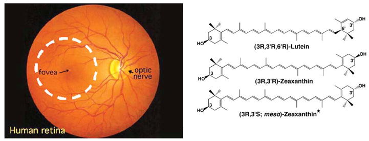
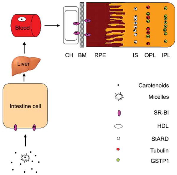
Two dietary carotenoids, lutein and zeaxanthin, are specifically delivered to the human macula at the highest concentration anywhere in the body. Whenever a tissue exhibits highly selective uptake of a compound, it is likely that one or more specific binding proteins are involved in the process. Over the past decade, our laboratory has identified and characterized several carotenoid-binding proteins from human retina including a pi isoform of glutathione S-transferase (GSTP1) as a zeaxanthin-binding protein, a member of the steroidogenic acute regulatory domain (StARD) family as a lutein-binding protein, and tubulin as a less specific, but higher capacity site for carotenoid deposition. In this article, we review the purification and characterization of these carotenoid-binding proteins, and we relate these ocular carotenoid-binding proteins to the transport and uptake role of serum lipoproteins and scavenger receptor proteins in a proposed pathway for macular pigment carotenoid delivery to the human retina.
Figures
References
-
- Home E. An account of the orifice in the retina of the human eye, discovered by Professor Soemmering: to which are added proofs of this appearance being extended to the eyes of other animals. Philos Trans R Soc Land. 1798;2:332–345.
-
- Wald G. Human vision and the spectrum. Science. 1945;101:653–658. - PubMed
-
- Bone RA, Laudrum JT, Tarsis SL. Preliminary identification of the human macular pigment. Vision Res. 1985;25:1531–1535. - PubMed
-
- Bone RA, Laudrum JT, Hime GW, Cains A, Zamor J. Stereochemistry of the human macular carotenoids. Inves Ophthalmol Vis Sci. 1993;34:2033–2040. - PubMed
-
- Whitehead AJ, Mares JA, Danis RP. Macular pigment. Arch Ophthalmol. 2006;124:1038–1045. - PubMed
Publication types
MeSH terms
Substances
Grants and funding
LinkOut - more resources
Full Text Sources
Other Literature Sources
Research Materials
Miscellaneous