. 2010 Sep;12(9):766-78.
doi: 10.1002/jgm.1496.

RH10 provides superior transgene expression in mice when compared with natural AAV serotypes for neonatal gene therapy

Affiliations

RH10 provides superior transgene expression in mice when compared with natural AAV serotypes for neonatal gene therapy

Chuhong Hu et al. J Gene Med. 2010 Sep.

Abstract

Background: Neonatal gene therapy is a promising strategy for treating diseases diagnosed before or shortly after birth. Early and long-term expression of therapeutic proteins may limit the consequences of genetic mutations and result in a potential 'cure'. Adeno-associated viral vectors have shown promise in many areas of adult gene therapy but their properties have not been systematically investigated in the neonate.

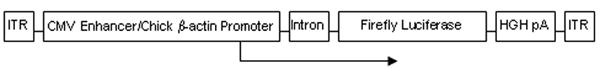
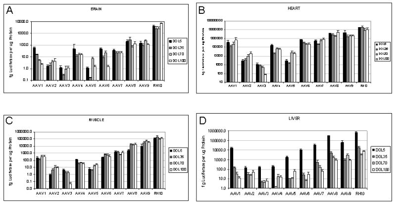
Methods: In these studies, using a constitutive promoter expressing luciferase, animals were administered one of ten serotypes of adeno-associated virus (AAV) on the second day of life. Examination of expression, organ growth and vector distribution, maintenance of expression and copy number were measured.

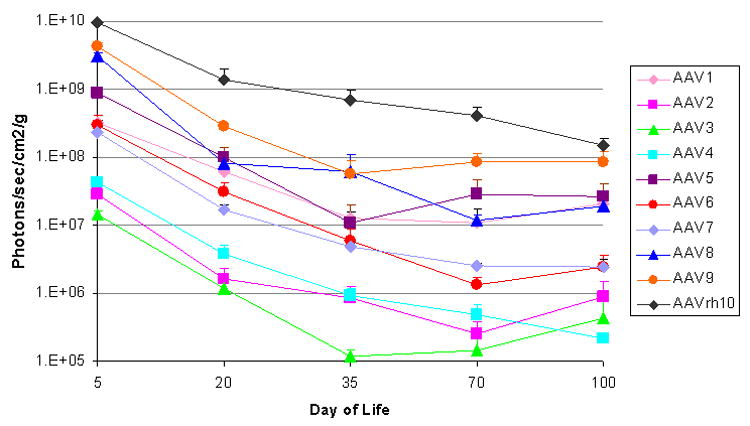
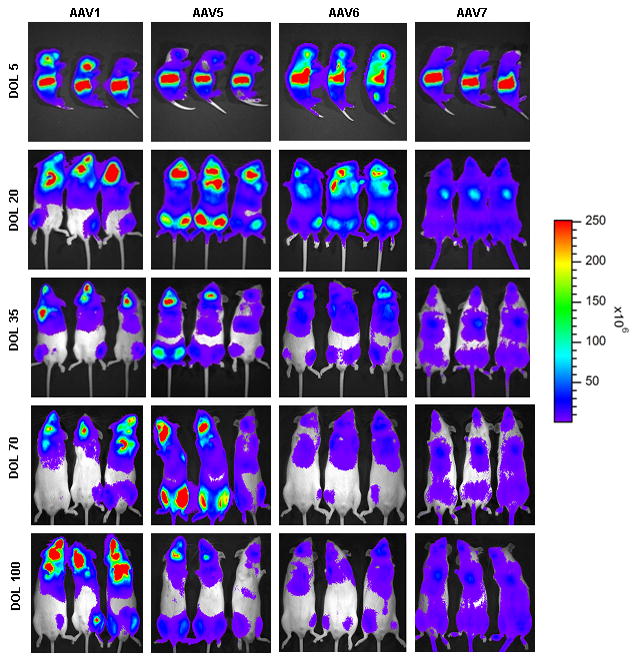
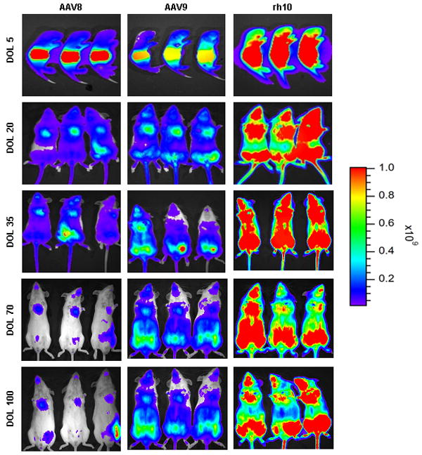
Results: All serotypes demonstrated expression and, in general, transduction of all organs within 3 days, albeit with different biodistribution patterns and expression levels. The highest expression was detected with AAVrh10, whereas the lowest was detected with AAV4. Expression and genomes declined with growth over the first 10 weeks of life; thereafter, to day 100, expression and genomes remained relatively stable. With the highest expressing vectors, whole animal expression at 100 days declined to approximately 10% of that detected on the fifth day. AAVrh10 maintained the highest expression level and copy number throughout these studies.

Conclusions: The impact of tissue and organ growth on the stability of AAV expression will be important if neonatal gene transfer is to be considered as a modality for human gene therapy. Although all vectors did demonstrate expression, rh10 holds the greater promise of the vectors tested to maintain copy number in both mitotic and post-mitotic tissues.


Figures

Figure 1
Figure 1
Diagram of recombinant adeno-associated viral vector (AAV.CB.Luciferase). AAV2 inverted terminal repeats (ITR) were used with pseudotyping to the respective capsid protein. The firefly luciferase cDNA is expressed under control of the cytomegalovirus (CMV) enhancer with chick β-actin promoter, β-globin intron, and with the human growth hormone (HGH) polyadenylation signal. The size of the AAV vector genome is 3.9 kb. The transgene was packaged with serotypes 1-rh10. Mice were injected intravenously on the second day of life with 2 × 10¹⁰ genome copies each.
Figure 2
Figure 2
Growth of mice after birth. Mice grow most rapidly during the first 5 weeks of life after which the rate of weight gain slows. During this period mouse weight increases 4-fold. Control, saline-injected mice of both genders were compared with AAV-injected mice for evidence of alteration or slower growth rates. (Data is expressed at mean ± SD. n=5 animals at each time point.)
Figure 3
Figure 3
Growth of selected organs during the first 100 days of life. A=brain; B=heart, lung; C=GI tract, liver, pancreas; D=kidney (n=5 animals at each time point). (Mean ± SD is presented.)
Figure 4
Figure 4
Dynamics of luciferase expression by bioluminescent imaging with animal growth. For each AAV serotype tested the mean total flux was calculated by averaging multiple animals at each time point (n=3-7 per time point). Mean ± SD of total flux emitted from the ventral surface was plotted.)
Figure 5
Figure 5
In vivo imaging of firefly luciferase after intravenous injection of AAV on the 2nd day of life demonstrates photon diffusion patterns. The images are shown at 5 days (72 hours after injection), 20 days, 35 days, 70 days, and 100 days of life. (A) Low-expressing group: Serotypes 2, 3, and 4 were classified as low-expressing in comparison to the other serotypes evaluated. Total flux in photons/second was acquired. (B) Medium-expressing group: Serotypes 1, 5, 6, 7 were classified as medium-expressing in comparison to other serotypes evaluated. (C) High-expressing group: Serotypes 8, 9, and rh10 were classified as high-expressing when compared with other serotypes. For all groups, images were acquired with the mice in the ventral position. Images within each group set with the same references such that side-by-side comparison can be made. Note changes in the legend of the pseudocolor scale per group. (n=3-7 animals per group at each time point.)
Figure 5
Figure 5
In vivo imaging of firefly luciferase after intravenous injection of AAV on the 2nd day of life demonstrates photon diffusion patterns. The images are shown at 5 days (72 hours after injection), 20 days, 35 days, 70 days, and 100 days of life. (A) Low-expressing group: Serotypes 2, 3, and 4 were classified as low-expressing in comparison to the other serotypes evaluated. Total flux in photons/second was acquired. (B) Medium-expressing group: Serotypes 1, 5, 6, 7 were classified as medium-expressing in comparison to other serotypes evaluated. (C) High-expressing group: Serotypes 8, 9, and rh10 were classified as high-expressing when compared with other serotypes. For all groups, images were acquired with the mice in the ventral position. Images within each group set with the same references such that side-by-side comparison can be made. Note changes in the legend of the pseudocolor scale per group. (n=3-7 animals per group at each time point.)
Figure 6
Figure 6
Luciferase expression profile in selected tissues over time from the panel of AAV serotypes (A=brain, B=heart, C=skeletal muscle, D=liver, E=intestine, F=pancreas, G=lung, H=kidney). The levels of luciferase protein in femtograms per microgram total protein were determined from mouse tissues after intravenous injection of AAV on the 2nd through 100 days of life. (Mean ± SEM is presented. N=5 animals at each time point.)
Figure 6
Figure 6
Luciferase expression profile in selected tissues over time from the panel of AAV serotypes (A=brain, B=heart, C=skeletal muscle, D=liver, E=intestine, F=pancreas, G=lung, H=kidney). The levels of luciferase protein in femtograms per microgram total protein were determined from mouse tissues after intravenous injection of AAV on the 2nd through 100 days of life. (Mean ± SEM is presented. N=5 animals at each time point.)

References

    1. Grimm D, Zhou S, Nakai H, Thomas CE, Storm TA, Fuess S, et al. Preclinical in vivo evaluation of pseudotyped adeno-associated virus vectors for liver gene therapy. Blood. 2003;102(7):2412–9. - PubMed
    1. Song Y, Lou HH, Boyer JL, Limberis MP, Vandenberghe LH, Hackett NR, et al. Functional cystic fibrosis transmembrane conductance regulator expression in cystic fibrosis airway epithelial cells by AAV6.2-mediated segmental trans-splicing. Hum Gene Ther. 2009;20(3):267–81. - PMC - PubMed
    1. Katakura S, Jennings K, Watanabe S, Adachi E, Thornton S, Gao GP, et al. Recombinant adeno-associated virus preferentially transduces human, compared to mouse, synovium: implications for arthritis therapy. Mod Rheumatol. 2004;14(1):18–24. - PubMed
    1. Simonelli F, Maguire AM, Testa F, Pierce EA, Mingozzi F, Bennicelli JL, et al. Gene therapy for Leber’s congenital amaurosis is safe and effective through 1.5 years after vector administration. Mol Ther. 2010;18(3):643–50. - PMC - PubMed
    1. Arruda VR, Stedman HH, Haurigot V, Buchlis G, Baila S, Favaro P, et al. Peripheral transvenular delivery of adeno-associated viral vectors to skeletal muscle as a novel therapy for hemophilia B. Blood. 2010 - PMC - PubMed

Publication types