. 2010 Oct 15;202(8):1287-95.
doi: 10.1086/656333.

Influenza enhances susceptibility to natural acquisition of and disease due to Streptococcus pneumoniae in ferrets

Affiliations

Influenza enhances susceptibility to natural acquisition of and disease due to Streptococcus pneumoniae in ferrets

Jonathan A McCullers et al. J Infect Dis. .

Abstract

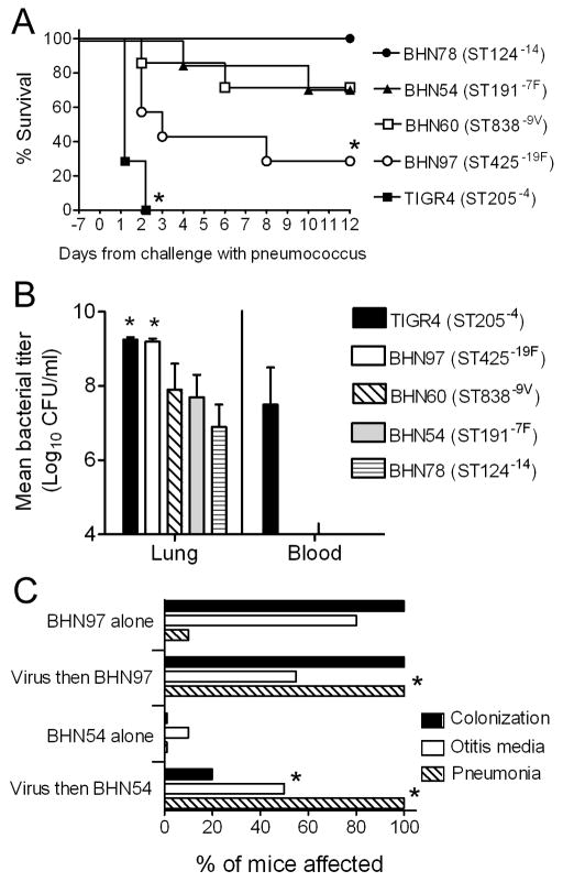
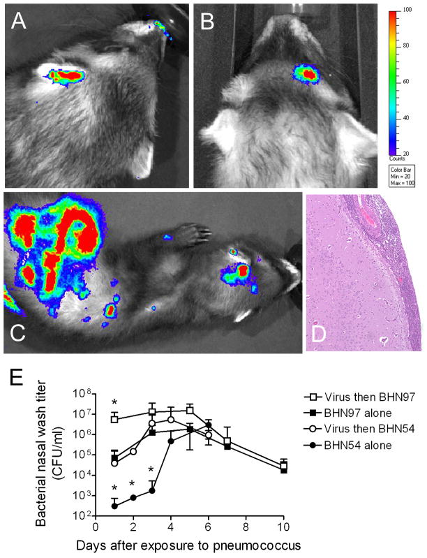
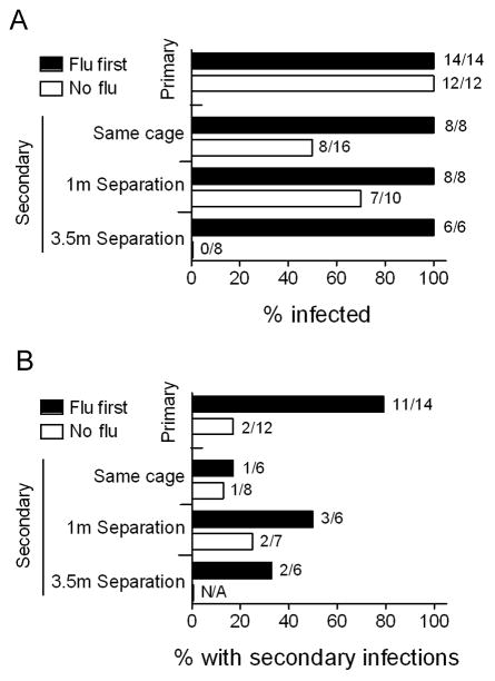
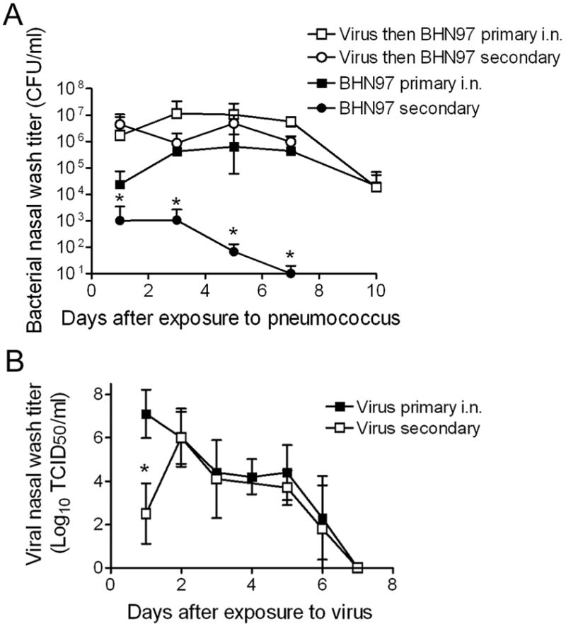
The role of respiratory viruses in the transmission of Streptococcus pneumoniae is poorly understood. Key questions, such as which serotypes are most fit for transmission and disease and whether influenza virus alters these parameters in a serotype-specific manner, have not been adequately studied. In a novel model of transmission in ferrets, we demonstrated that pneumococcal transmission and disease were enhanced if donors had previously been infected with influenza virus. Bacterial titers in nasal wash, the incidence of mucosal and invasive disease, and the percentage of contacts that were infected all increased. In contact ferrets, viral infection increased their susceptibility to S. pneumoniae acquisition both in terms of the percentage infected and the distance over which they could acquire infection. These influenza-mediated effects on colonization, transmission, and disease were dependent on the pneumococcal strain. Overall, these data argue that the relationship between respiratory viral infections, acquisition of pneumococci, and development of disease in humans needs further study to be better understood.


Conflict of interest statement

The authors declare no conflicts of interest exist.

Figures

Figure 1
Figure 1. The impact of influenza virus on disease differs with pneumococcal strain used
A) Groups of 6 mice were infected intranasally with 30 TCID50 of influenza virus PR8 7 days prior to challenge with 1×10⁵ CFU of S. pneumoniae strains BHN78 (type 14), BHN54 (type 7F), BHN60 (type 9V), BHN97 (type 19F), or TIGR4 (type 4) and followed for mortality. An asterisk (*) indicates a significant difference (p < 0.05) by log-rank test on the Kaplan-Meier survival data vs. the other groups. B) Lung and blood titers were taken 48 hours after secondary challenge from groups of 4 mice infected as above. An asterisk (*) indicates a significant difference (p < 0.05) by ANOVA vs. the BHN78, BHN54, and BHN60 groups. C) Groups of 5 mice were infected intranasally with 30 TCID50 of influenza virus PR8 or were mock-infected with PBS 7 days prior to challenge with 1×10⁵ CFU of S. pneumoniae using versions of BHN54 and BHN97 engineered to express luciferase, and the incidence of rhinitis, otitis media, and pneumonia as determined by bioluminescent imaging were determined over a 14 day period. An asterisk (*) indicates a significant difference (p < 0.05) by Student’s t-test with Bonferroni correction vs. the corresponding bacteria alone group.
Figure 2
Figure 2. Influenza infection predisposes ferrets to secondary pneumococcal infections
Ferrets infected with 1×10⁵ TCID50 of influenza virus A/Sydney/5/97 (H3N2) and challenged 3 days later with 1×10⁷ CFU of pneumococcal strain BHN97 were assessed by bioluminescent imaging for foci of bacteria representing sites of secondary bacterial infections. Representative images from ferrets with A) otitis media, B) sinusitis, and C) disseminated disease are pictured. The scale indicates the relative light units per pixel. The ferret in C) had D) meningitis characterized by expansion of the meninges by exuberant infiltrates of neutrophils with admixed macrophages, lymphocytes and necrotic cellular debris. Inflammation was confined to the meninges; however there was a mild response on the superficial brain surface consisting of edema and gliosis. E) Groups of 5 ferrets were infected with influenza or were mock-infected with PBS 3 days prior to challenge with pneumococcal strains BHN54 or BHN97. An asterisk (*) indicates a significant difference by ANOVA in nasal wash titer compared to all other groups at that timepoint (p < 0.05).
Figure 3
Figure 3. Influenza virus enhances transmission between ferrets
A summary of several experiments (from Table 1) is presented demonstrating the percentage of ferrets A) infected or B) which developed secondary bacterial infections with S. pneumoniae strain BHN97 (considering only those which were shedding), stratified by whether the ferrets were directly infected by inoculation under anesthesia (primary) or infected naturally by transmission (secondary), by whether they were previously infected with influenza virus (flu first) or were naïve to influenza virus at the time of infection or exposure (no flu), and, for contact ferrets, by physical proximity to the donor ferrets (same cage, 1 meter of separation, 3.5 meters of separation). N/A indicates no ferrets were shedding.
Figure 4
Figure 4. Bacterial and viral titers in nasal washes are similar in singly infected compared to co-infected ferrets
A summary of several experiments is presented demonstrating A) mean bacterial titers in nasal washes from ferrets directly infected with S. pneumoniae strain BHN97 by inoculation under anesthesia (primary i.n.; n = 8 per group) or infected naturally by transmission (secondary; n = 15 per group) stratified by whether they were previously infected with influenza virus or were naive to influenza virus at the time of infection or exposure, and B) mean viral titers in ferrets directly infected with influenza virus by inoculation under anesthesia (primary i.n.; n = 13 per group) or infected naturally by transmission (secondary; n = 4 per group). An asterisk (*) indicates a significant difference (p < 0.05) in titer compared to all other groups at that time point.
Figure 5
Figure 5. Presence or absence of accessory regions (ARs) and virulence genes in strains used in this study
A) Presence or absence of ARs were determined for strains utilized in the study including TIGR4. Yellow, AR is present. White, some genes from the AR are present and some are absent. Dark blue, AR is absent. ARs are defined in [10]. B) Presence or absence of specific genes linked to invasion by signature tagged mutagenesis [10] were determined for strains used in the study compared to known loci in TIGR4 and R6. Yellow, gene is present. Light blue, gene is likely to be absent but the p value is not significant. Dark blue, gene is absent (p < 0.01). Gray, there are no data or the data are unclear from this analysis.

Comment in

References

    1. Bridy-Pappas AE, Margolis MB, Center KJ, Isaacman DJ. Streptococcus pneumoniae: description of the pathogen, disease epidemiology, treatment, and prevention. Pharmacotherapy. 2005;25(9):1193–212. - PubMed
    1. De Wals P, Erickson L, Poirier B, Pepin J, Pichichero ME. How to compare the efficacy of conjugate vaccines to prevent acute otitis media? Vaccine. 2009;27(21):2877–83. - PubMed
    1. McCullers JA. Insights into the interaction between influenza virus and pneumococcus. Clin Microbiol Rev. 2006;19(3):571–82. - PMC - PubMed
    1. Peltola VT, McCullers JA. Respiratory viruses predisposing to bacterial infections: role of neuraminidase. Pediatr Infect Dis J. 2004;23(1 Suppl):S87–S97. - PubMed
    1. Finland M. Recent advances in the epidemiology of pneumococcal infections. Medicine (Baltimore) 1942;21(3):307–44.

Publication types