. 2010 Sep 7:6:51.
doi: 10.1186/1744-8069-6-51.

Inhibition of class II histone deacetylases in the spinal cord attenuates inflammatory hyperalgesia

Affiliations

Inhibition of class II histone deacetylases in the spinal cord attenuates inflammatory hyperalgesia

Guang Bai et al. Mol Pain. .

Abstract

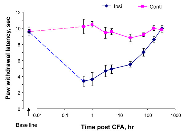
Background: Several classes of histone deacetylases (HDACs) are expressed in the spinal cord that is a critical structure of the nociceptive pathway. HDAC-regulated histone acetylation is an important component of chromatin remodeling leading to epigenetic regulation of gene transcription. To understand the role of histone acetylation in epigenetic regulation of pathological pain, we have studied the impact of different classes of HDACs in the spinal cord on inflammatory hyperalgesia induced by complete Freund's adjuvant (CFA).

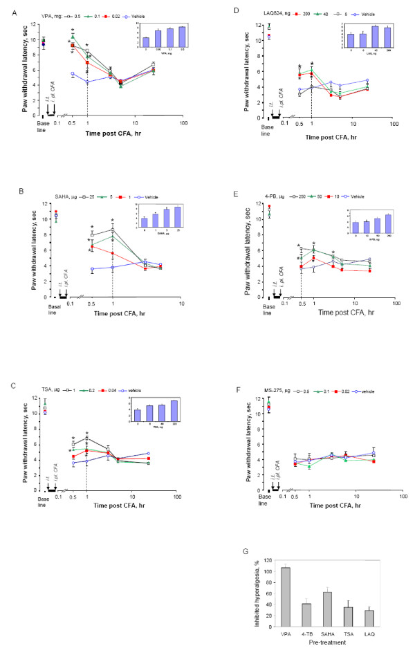
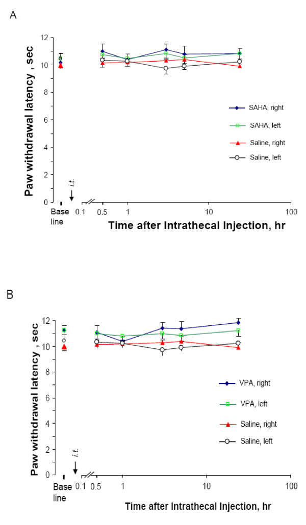
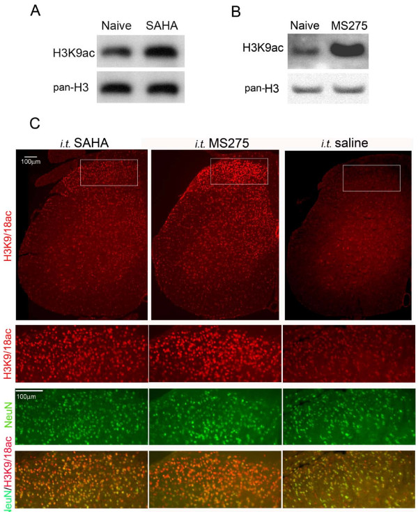
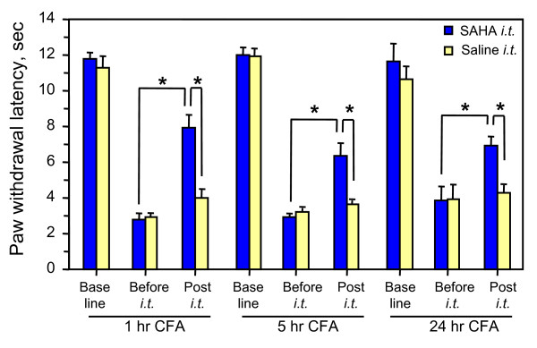
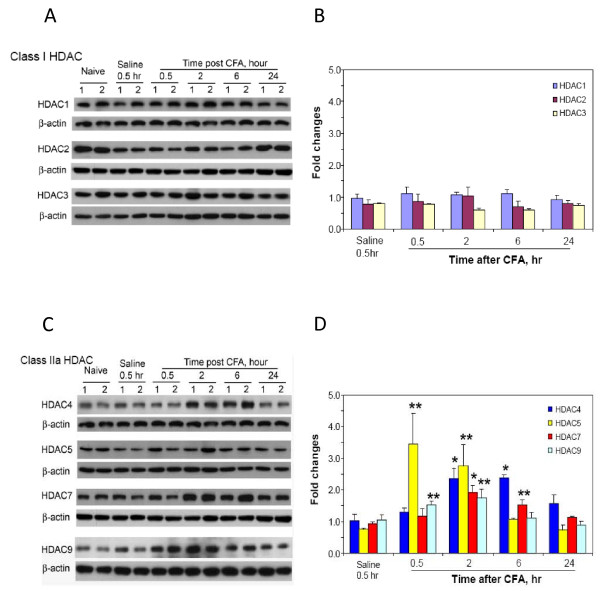
Results: We intrathecally applied inhibitors specific to different classes of HDACs and evaluated their impact on inflammatory hyperalgesia. Pre-injected inhibitors targeting class I as well as II (SAHA, TSA, LAQ824) or IIa (VPA, 4-PB) HDACs significantly delayed the thermal hyperalgesia induced by unilateral CFA injection in the hindpaw. Existing hyperalgesia induced by CFA was also attenuated by the HDAC inhibitors (HDACIs). In contrast, these inhibitors did not interfere with the thermal response either in naïve animals, or on the contralateral side of inflamed animals. Interestingly, MS-275 that specifically inhibits class I HDACs failed to alter the hyperalgesia although it increased histone 3 acetylation in the spinal cord as SAHA did. Using immunoblot analysis, we further found that the levels of class IIa HDAC members (HDAC4, 5, 7, 9) in the spinal dorsal horn were upregulated following CFA injection while those of class I HDAC members (HDAC1, 2, 3) remained stable or were slightly reduced.

Conclusions: Our data suggest that activity of class II HDACs in the spinal cord is critical to the induction and maintenance of inflammatory hyperalgesia induced by CFA, while activity of class I HDACs may be unnecessary. Comparison of the effects of HDACIs specific to class II and IIa as well as the expression pattern of different HDACs in the spinal cord in response to CFA suggests that the members of class IIa HDACs may be potential targets for attenuating persistent inflammatory pain.


Figures

Figure 1
Figure 1
Thermal hyperalgesia induced by CFA in mice. Paw withdrawal latency of mice to a thermal stimulus on hindpaws were measured from both hind paws before and after unilateral CFA injection. Mean ± s.e. values from 10 animals were presented. Contl, contralateral side; Ipsi, ipsilateral side.
Figure 2
Figure 2
Short-term attenuation effects of pre-injected HDACI on thermal hyperalgesia in mice. Mice received intrathecally injected vehicle or HDACI at indicated dose 30 min before unilateral injection of CFA. Paw withdrawal latency was measured before injections as baseline, and after CFA injections as hyperalgesia response. Following HDACIs were injected for panel (in parenthesis) VPA (A), 4-BP (B), SAHA (C), TSA (D), LAQ824 (E). The inserted bar graphs showed dose dependence from the time lined up with a dash line. Mean values of eight animals per group plus standard errors are presented for the responses on the ipsilateral side. * P < 0.01, # P < 0.05: compared to vehicle at the same time point. G. Comparison of inhibition of thermal hyperalgesia by tested HDACI. Inhibition of hyperalgesia by all inhibitors at the maximal dose tested 30 min after CFA injection were calculated as described in method. One-way ANOVA analysis indicated there was no significant difference among HDAC-specific inhibitors, but all of them showed significant difference in comparison to VPA (P < 0.05).
Figure 3
Figure 3
Effects of HDACI on thermal nociception on naïve mice. Naïve mice treated with i.t. SAHA (25 μg) or VPA (0.5 mg) were tested for their hindpaw response to a noxious heat beam for indicated time. Mean values + s.e. of PWL from 8 animals each group were presented. No significance was revealed for comparison among tested period either at the ipsilateral or contralateral side.
Figure 4
Figure 4
Histone acetylation in the spinal cord after HDACI treatment. Histone acetylation in the lumbar spinal cord of mice receiving i.t. SAHA (25 μg) or MS-275 (0.5 μg) for 30 min was analyzed by immunoblot (A, B) and immunofluorescent histochemistry (C) for antigens indicated. Animals receiving i.t. saline were used as control. Images of the H3K9/18ac signals in the left half of the lumbar spinal cord are shown in the first row in C. Immunosignals of indicated antigens in the superficial dorsal horn are presented in the rest rows in C.
Figure 5
Figure 5
Attenuation of existing thermal hyperalgesia by HDACI. Mice were treated with CFA i.pl. for indicated time and then intrathecally injected with 25 μg SAHA for 30 min before measurement of thermal response. Control animals were treated with i.t. saline after CFA injection for the same time. Basal line was measured before the CFA injection. Student t-test was used to examine the significance as indicated groups. * P < 0.01.
Figure 6
Figure 6
Changes of HDACs' levels in the dorsal spinal cord after CFA injection into the hindpaw. The levels of HDACs in the dorsal spinal cord were analyzed by immunoblot using antibodies specific to each target as indicated. Examples of blots are shown in A and C. Digitally analyzed levels of each HDAC after normalization to β-actin are shown in B for class I HDACs and in D for class IIa HDACs. Mean values plus s.e. are averaged from 4 to 6 animals. Student t-test was used to examine the significance as indicated groups. * P < 0.01; ** P < 0.05.

References

    1. LaCroix-Fralish ML, Ledoux JB, Mogil JS. The Pain Genes Database: An interactive web browser of pain-related transgenic knockout studies. Pain. 2007;131:3.e1–3.e4. doi: 10.1016/j.pain.2007.04.041. - DOI - PubMed
    1. Geranton SM, Morenilla-Palao C, Hunt SP. A role for transcriptional repressor methyl-CpG-binding protein 2 and plasticity-related gene serum- and glucocorticoid-inducible kinase 1 in the induction of inflammatory pain states. J Neurosci. 2007;27:6163–6173. doi: 10.1523/JNEUROSCI.1306-07.2007. - DOI - PMC - PubMed
    1. Griffin RS, Costigan M, Brenner GJ, Ma CH, Scholz J, Moss A, Allchorne AJ, Stahl GL, Woolf CJ. Complement induction in spinal cord microglia results in anaphylatoxin C5a-mediated pain hypersensitivity. J Neurosci. 2007;27:8699–8708. doi: 10.1523/JNEUROSCI.2018-07.2007. - DOI - PMC - PubMed
    1. Rohl T, Kurreck J. RNA interference in pain research. J Neurochem. 2006;99:371–380. doi: 10.1111/j.1471-4159.2006.04082.x. - DOI - PubMed
    1. Cheng HY, Pitcher GM, Laviolette SR, Whishaw IQ, Tong KI, Kockeritz LK, Wada T, Joza NA, Crackower M, Goncalves J, Sarosi I, Woodgett JR, Oliveira-dos-Santos AJ, Ikura M, van der Kooy D, Salter MW, Penninger JM. DREAM is a critical transcriptional repressor for pain modulation. Cell. 2002;108:31–43. doi: 10.1016/S0092-8674(01)00629-8. - DOI - PubMed

Publication types

MeSH terms