. 2011 Jan;39(1):225-34.
doi: 10.1093/nar/gkq769. Epub 2010 Sep 7.

Allotopic expression of mitochondrial-encoded genes in mammals: achieved goal, undemonstrated mechanism or impossible task?

Affiliations

Allotopic expression of mitochondrial-encoded genes in mammals: achieved goal, undemonstrated mechanism or impossible task?

Ester Perales-Clemente et al. Nucleic Acids Res. 2011 Jan.

Abstract

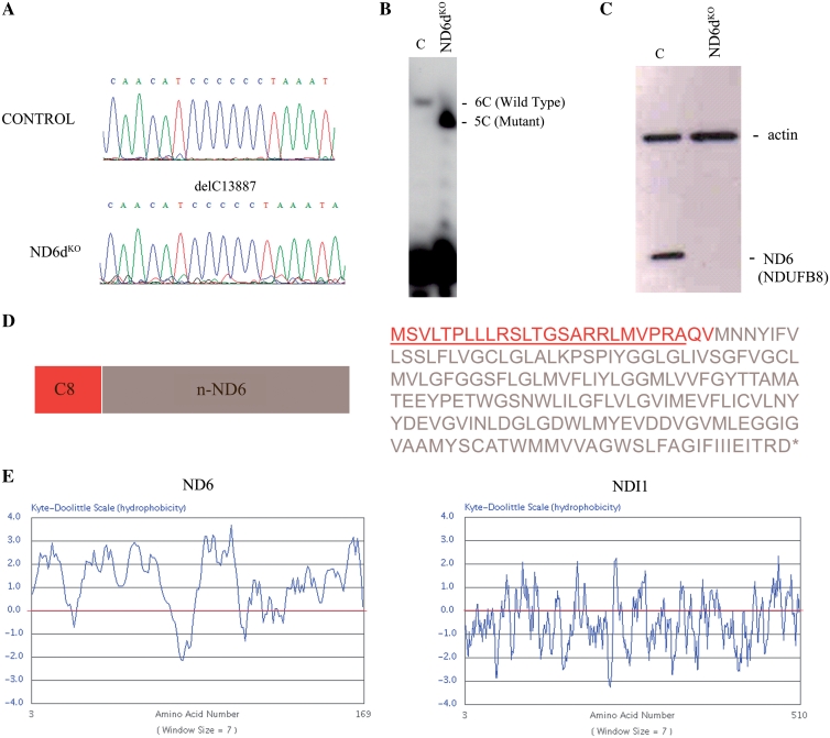
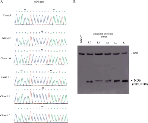
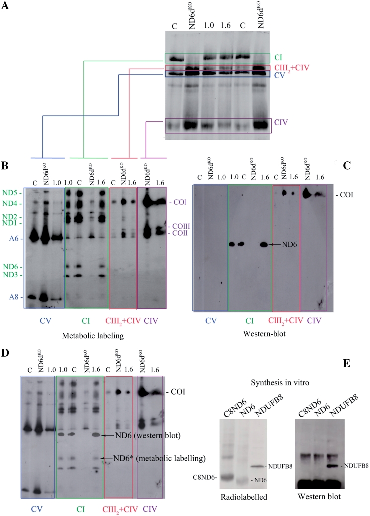
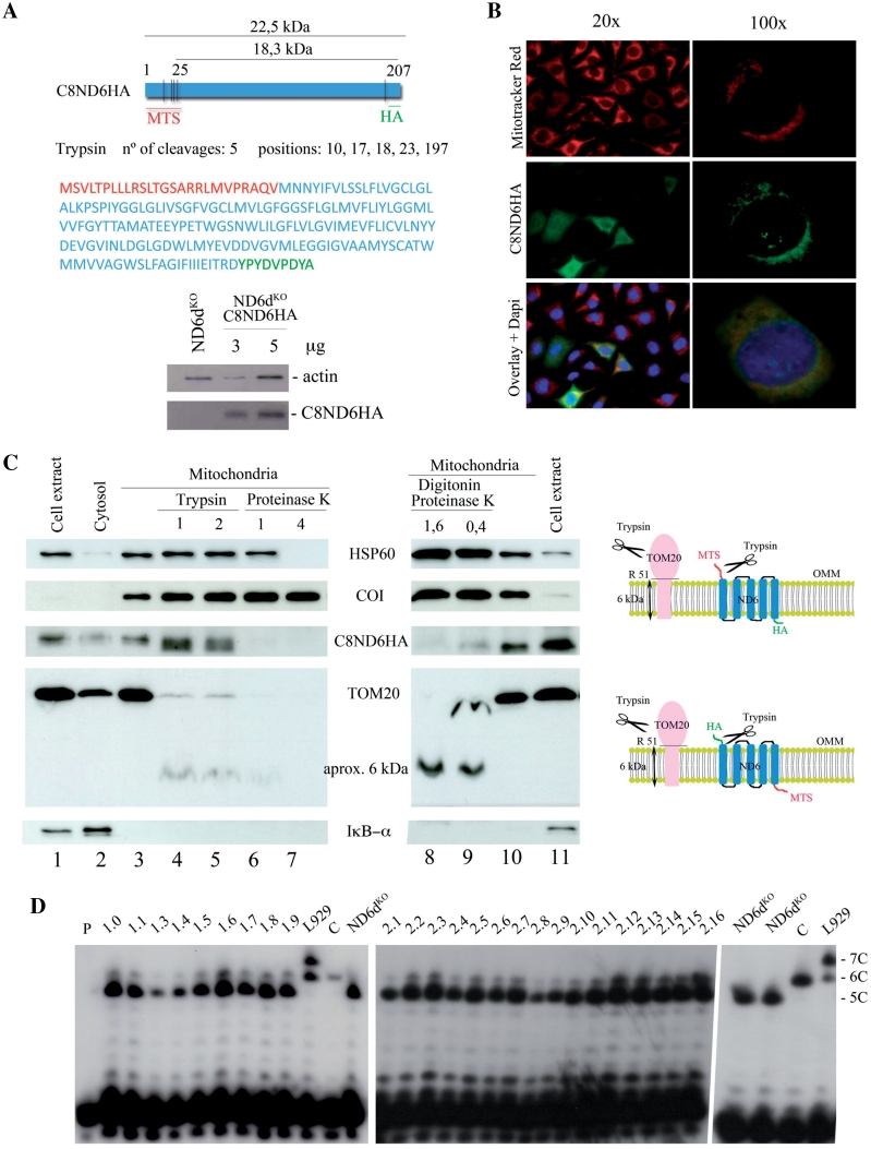
Mitochondrial-DNA diseases have no effective treatments. Allotopic expression-synthesis of a wild-type version of the mutated protein in the nuclear-cytosolic compartment and its importation into mitochondria-has been proposed as a gene-therapy approach. Allotopic expression has been successfully demonstrated in yeast, but in mammalian mitochondria results are contradictory. The evidence available is based on partial phenotype rescue, not on the incorporation of a functional protein into mitochondria. Here, we show that reliance on partial rescue alone can lead to a false conclusion of successful allotopic expression. We recoded mitochondrial mt-Nd6 to the universal genetic code, and added the N-terminal mitochondrial-targeting sequence of cytochrome c oxidase VIII (C8) and the HA epitope (C8Nd6HA). The protein apparently co-localized with mitochondria, but a significant part of it seemed to be located outside mitochondria. Complex I activity and assembly was restored, suggesting successful allotopic expression. However, careful examination of transfected cells showed that the allotopically-expressed protein was not internalized in mitochondria and that the selected clones were in fact revertants for the mt-Nd6 mutation. These findings demonstrate the need for extreme caution in the interpretation of functional rescue experiments and for clear-cut controls to demonstrate true rescue of mitochondrial function by allotopic expression.


Figures

Figure 1.
Figure 1.
Allotopic expression strategy. (A) Chromatograms showing the homoplasmic mutations found in the mt-Nd6 gene. (B) Allele-specific termination of primer extension assay to confirm the homoplasmy of the deletion of one C in ND6dKO cells. (C) Western blot of total protein from control and mutant cells with the anti-ND6 antibody marketed by Molecular Probes (which in fact recognizes NDUFB8; see text and Figure 3). Actin expression was probed as a loading control. (D) Amino acid sequence and map of C8ND6 (the mt-Nd6 gene sequence recoded to the universal genetic code using Backtranslation software). Red letters correspond to the C8 MTS sequence plus the first two amino acids of mature COX8 (QV). (E) Kyte and Doolittle plots illustrating the highly hydrophobic character of the mitochondrial ND6 protein, compared with the yeast protein NDI1.
Figure 2.
Figure 2.
Analysis of galactose-resistant clones. (A) Sequence chromatograms showing conservation of the 13887 delC mutation in the mt-Nd6 gene in four clones (1.0, 1.1, 1.6 and 1.7) obtained after galactose selection. (B) Western blot of total protein from wild-type (C), ND6dKO cells and galactose-resistant clones allotopically expressing C8ND6 protein. The blot was probed with the anti-ND6 from Molecular Probes, which in fact recognizes NDUFB8 (see text and Figure 3) and with anti-actin as a loading control.
Figure 3.
Figure 3.
Galactose-resistant ND6dKOC8ND6 clones express wild-type mt-Nd6. (A) Metabolic labelling of assembled OXPHOS complexes. Fluorogram after Blue-Native gel electrophoresis of the mitochondrial translation products of wild-type (C), mutant (ND6dKO) and two galactose-resistant ND6dKOC8ND6 clones (1.0 and 1.6). Before protein extraction, cells were pulse-labelled with [35S]-methionine for 1 h in the presence of cycloheximide and chased for 12 h. (B). Fluorogram of mitochondrial translation products separated by 2D electrophoresis. Isolated OXPHOS complexes obtained in the first dimension were resolved on denaturing gels and electro-transferred to PVDF membrane as indicated. (C) Western blot of the second dimension gel with the Molecular Probes anti-ND6 antibody (complex I) and anti-CO1 (complex IV). (D). Superposition of the fluorogram and western blot from B and C, showing that the anti-ND6 antibody does not recognize ND6. (E) Identification of the specificity of the anti-ND6 antibody. C8ND6, ND6 and NDUFB8, which participates in the assembly of mitochondrial complex I, were synthesized in vitro in the presence of [35S] methionine. The left panel shows an autoradiogram of a PVDF membrane blotted from a 12% SDS–polyacrylamide gel. The right panel shows a western blot of the membrane with the Molecular Probes anti-ND6 antibody.
Figure 4.
Figure 4.
Allotopically expressed ND6 is not incorporated into the inner mitochondrial membrane. (A) Upper panel: map and amino acid sequence of C8ND6HA. The COX8 MTS is shown in red, the recoded mt-Nd6 sequence in blue and the HA epitope in green. Lower panel: western blot showing expression of C8ND6HA in lentivirally transformed ND6dKO cells. The blot was probed with anti-HA (Roche) and with anti-actin as a loading control. (B) Subcellular localization of C8ND6HA in transformed cells. ND6kdKOC8ND6HA cells were visualized by indirect inmunofluorescence using anti-HA antibody and Mitotracker® Red (Invitrogen). The precursor polypeptide was expressed and the protein apparently co-localized with mitochondria. (C) Left panel: western blot of isolated mitochondria from ND6dKOC8ND6HA cells, showing that allotopically expressed C8ND6HA is not internalized. Lanes (from left to right): 1, cell extract; 2, cytosol; 3, untreated isolated mitochondria; 4 and 5, mitochondria treated with 1 or 2 µg trypsin/100 µg mitochondrial protein; 6 and 7, mitochondria treated with 1 or 4 µg proteinase K/100 µg mitochondria; 8 and 9, mitochondria treated with 1.6 or 0.4 µg proteinase K/100 µg mitochondria plus 0.15 mg/ml digitonin; 10, isolated mitochondria and 11, cell extract. The protein loading was 20 µg for lanes 1, 2 and 11; 10 µg for lanes 3–7 and 10 and 50 µg for lanes 8 and 9. Right panel: diagram showing the likely actions of trypsin on TOM20 and allotopically expressed C8ND6HA proteins. (D) Allele-specific primer extension termination analysis. All the clones obtained after galactose selection were revertants for the mt-Nd6 mutation, in both experiments (C8ND6 and C8ND6HA expression). P: primer; 1.0–1.9, galactose-resistant clones obtained after allotopic expression of C8ND6; L929, mouse cell line heteroplasmic for the 13887iC mutation in mt-ND6 (25); 2.1-2.16, galactose-resistant clones obtained after allotopic expression of C8ND6HA; C, wild-type cells.

References

    1. Woischnik M, Moraes CT. Pattern of organization of human mitochondrial pseudogenes in the nuclear genome. Genome Res. 2002;12:885–893. - PMC - PubMed
    1. Saccone C, Gissi C, Reyes A, Larizza A, Sbisa E, Pesole G. Mitochondrial DNA in metazoa: degree of freedom in a frozen event. Gene. 2002;286:3–12. - PubMed
    1. Wallace DC. Why do we still have a maternally inherited mitochondrial DNA? Insights from evolutionary medicine. Annu. Rev. Biochem. 2007;76:781–821. - PubMed
    1. Wallace DC. Structure and evolution of organelle genomes. Microbiol. Rev. 1982;46:208–240. - PMC - PubMed
    1. von Heijne G. Why mitochondria need a genome. FEBS Lett. 1986;198:1–4. - PubMed

Publication types