. 2010 Dec;95(12):2013-21.
doi: 10.3324/haematol.2010.029827. Epub 2010 Sep 7.

Histone deacetylase 2 is required for chromatin condensation and subsequent enucleation of cultured mouse fetal erythroblasts

Affiliations

Histone deacetylase 2 is required for chromatin condensation and subsequent enucleation of cultured mouse fetal erythroblasts

Peng Ji et al. Haematologica. 2010 Dec.

Abstract

Background: During the final stages of differentiation of mammalian erythroid cells, the chromatin is condensed and enucleated. We previously reported that Rac GTPases and their downstream target, mammalian homolog of Drosophila diaphanous 2 (mDia2), are required for enucleation of in vitro cultured mouse fetal liver erythroblasts. However, it is not clear how chromatin condensation is achieved and whether it is required for enucleation.

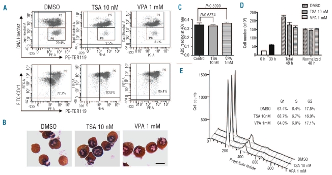
Design and methods: Mouse fetal liver erythroblasts were purified from embryonic day 14.5 pregnant mice and cultured in erythropoietin-containing medium. Enucleation was determined by flow-cytometry based analysis after treatment with histone deacetylase inhibitors or infection with lentiviral short hairpin RNA.

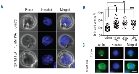
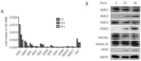
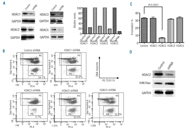
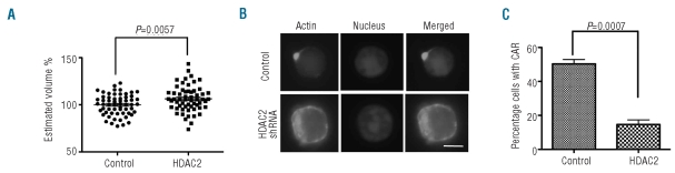
Results: We showed that histone deacetylases play critical roles in chromatin condensation and enucleation in cultured mouse fetal liver erythroblasts. Enzymatic inhibition of histone deacetylases by trichostatin A or valproic acid prior to the start of enucleation blocked chromatin condensation, contractile actin ring formation and enucleation. We further demonstrated that histone deacetylases 1, 2, 3 and 5 are highly expressed in mouse fetal erythroblasts. Short hairpin RNA down-regulation of histone deacetylase 2, but not of the other histone deacetylases, phenotypically mimicked the effect of trichostatin A or valproic acid treatment, causing significant inhibition of chromatin condensation and enucleation. Importantly, knock-down of histone deacetylase 2 did not affect erythroblast proliferation, differentiation, or apoptosis.

Conclusions: These results identify histone deacetylase 2 as an important regulator, mediating chromatin condensation and enucleation in the final stages of mammalian erythropoiesis.


Figures

Figure 1.
Figure 1.
Histone deacetylases are required for enucleation, but not terminal differentiation or proliferation, of cultured mouse fetal liver erythroblasts. (A) Purified TER119-negative E13.5 mouse fetal liver erythroid progenitor cells were cultured in vitro in fibronectin-coated plates in medium containing serum and erythropoietin (Epo). Tricostatin A 10 nM or valproic acid (VPA) 1 mM was added after 30 h in culture and cells were harvested after 48 h. The enucleation and differentiation status of cultured cells after TSA or VPA treatment were analyzed by flow cytometric analysis using staining with Hoechst 33342 and TER119–PE, and FITC–CD71 and TER119–PE, respectively. The percentages of enucleated incipient reticulocytes (top panels) and TER119-positive cells (bottom panels) are illustrated. (B) The same cells as in A were stained with benzidine-Giemsa. The scale bar represents 7 μm. (C) Hemoglobin concentrations of indicated cells analyzed at 48 h in culture. (D) The same cells as in A were counted at 0, 30 and 48 h in culture and the total number is shown. The total number of cells at 48 h was normalized, calculated as the number of nucleated erythroblasts plus one half of the total numbers of reticulocytes and nuclei. (E) Cell cycle analysis of the indicated cells at 40 h in culture. The percentages of the cells at G1, S, and G2 phases are shown.
Figure 2.
Figure 2.
Histone deacetylases are required for chromatin condensation of cultured mouse fetal liver erythroblasts. (A) Confocal microscopy analysis of TSA-treated mouse fetal liver erythroblasts. TER119-negative mouse fetal liver erythroblasts were cultured as in Figure 1. After 30 h, the indicated amount of TSA was added. Cells were harvested after 42 h and analyzed by confocal microscopy. Z-projections of DNA (blue) are shown. The scale bar represents 5 μm (B) Quantification of the estimated nuclear volume of the cells shown in A and VPA-treated cells. *P<0.001, **P<0.0001. (C) The same cells as in A treated with 10 nM of TSA after 30 h in culture were harvested after 48 h and stained with Alexa Fluor 488–phalloidin. The scale bar represents 8 μm.
Figure 3.
Figure 3.
HDAC 1, 2, 3 and 5 are highly expressed in cultured mouse fetal liver erythroblasts. (A) The mRNA expression levels of different HDAC during in vitro culture of TER119-negative mouse fetal erythroblasts, as quantified by quantitative real-time polymerase chain reaction analysis. The relative level compared to 18S rRNA was calculated using the ΔΔCt method. (B) Protein expression levels of HDAC 1, 2, 3 and 5, as well as histone H4 and acetylated H4K16 during in vitro culture of TER119-negative mouse fetal erythroblasts analyzed by western blot. The mDia2 and GAPDH protein levels are shown as positive and internal loading controls, respectively.
Figure 4.
Figure 4.
Depletion of HDAC2 inhibits enucleation and chromatin condensation (A) Depletion of HDAC 1, 2, 3 and 5 in primary erythroid cells by shRNA. TER119-negative mouse fetal erythroblasts were infected with lentiviral vectors encoding shRNA specific for HDAC 1, 2, 3 or 5 and cultured for 48 h. Lysates were subjected to western blotting for individual proteins as indicated. Controls show GAPDH levels. The gels were scanned and quantified. Controls were set as 100. (B) Flow cytometric analysis of cells infected with lentirviruses encoding HDAC 1, 2, 3 or 5 shRNA and cultured for 2 days, as in Figure 1. Cells were stained with TER119–PE and Hoechst 33342 and analyzed by FACS. The percentages of incipient reticulocytes are indicated. (C) Quantification of incipient reticulocytes in cells infected with indicated lentiviruses. (D) Western blot analysis of acetylated H4K16 when HDAC2 was down-regulated by shRNA.
Figure 5.
Figure 5.
Depletion of HDAC2 blocks chromatin condensation and contractive actin ring (CAR) formation of cultured mouse fetal liver erythroblasts. (A) Quantification of the estimated nuclear volume of 2-day cultured mouse fetal liver erythroblasts infected with lentiviruses expressing HDAC2 shRNA. (B) Alexa Fluor 488–phalloidin staining the same cells as in A. The scale bar represents 5 μm. (C). Quantification from B of cells containing a CAR. Three independent experiments were performed with roughly 200 cells counted in randomly picked areas.
Figure 6.
Figure 6.
Depletion of HDAC2 does not block terminal differentiation and proliferation of cultured mouse fetal liver erythroblasts. (A) Flow cytometric analysis of cells infected with lentivirus encoding HDAC1, HDAC2, HDAC3 or HDAC5 shRNA and cultured for 2 days, as in Figure 4B. Cells were stained with TER119–PE and FITC-CD71 and analyzed by FACS. The percentages of TER119-positive cells are quantified. (B) Quantification of TER119-positive cells from A. (C) Benzidine-Giemsa staining of cells 48 h after infection with lentivirus encoding control or HDAC2 shRNA. The scale bar represents 8 μm. (D) Hemoglobin concentration of cells 48 h after infection with lentivirus encoding control or HDAC2 shRNA. (C) The same cells as in A were counted after 48 h in culture. The total number and the normalized number, as in Figure 1D, are shown.

Comment in

  • Erythroblast enucleation.
    Migliaccio AR. Migliaccio AR. Haematologica. 2010 Dec;95(12):1985-8. doi: 10.3324/haematol.2010.033225. Haematologica. 2010. PMID: 21123437 Free PMC article. No abstract available.

References

    1. Richmond TD, Chohan M, Barber DL. Turning cells red: signal transduction mediated by erythropoietin. Trends Cell Biol. 2005;15(3):146–55. - PubMed
    1. Ji P, Jayapal SR, Lodish HF. Enucleation of cultured mouse fetal erythroblasts requires Rac GTPases and mDia2. Nat Cell Biol. 2008;10(3):314–21. - PubMed
    1. Yoshida H, Kawane K, Koike M, Mori Y, Uchiyama Y, Nagata S. Phosphatidylserine-dependent engulfment by macrophages of nuclei from erythroid precursor cells. Nature. 2005;437(7059):754–8. - PubMed
    1. Koury ST, Koury MJ, Bondurant MC. Cytoskeletal distribution and function during the maturation and enucleation of mammalian erythroblasts. J Cell Biol. 1989;109(6 Pt 1):3005–13. - PMC - PubMed
    1. Chasis JA, Prenant M, Leung A, Mohandas N. Membrane assembly and remodeling during reticulocyte maturation. Blood. 1989;74(3):1112–20. - PubMed

Publication types

MeSH terms