. 2010 Sep 15;70(18):7283-93.
doi: 10.1158/0008-5472.CAN-09-4125. Epub 2010 Sep 7.

Angiopoietin-4 promotes glioblastoma progression by enhancing tumor cell viability and angiogenesis

Affiliations

Angiopoietin-4 promotes glioblastoma progression by enhancing tumor cell viability and angiogenesis

Melissa K Brunckhorst et al. Cancer Res. .

Abstract

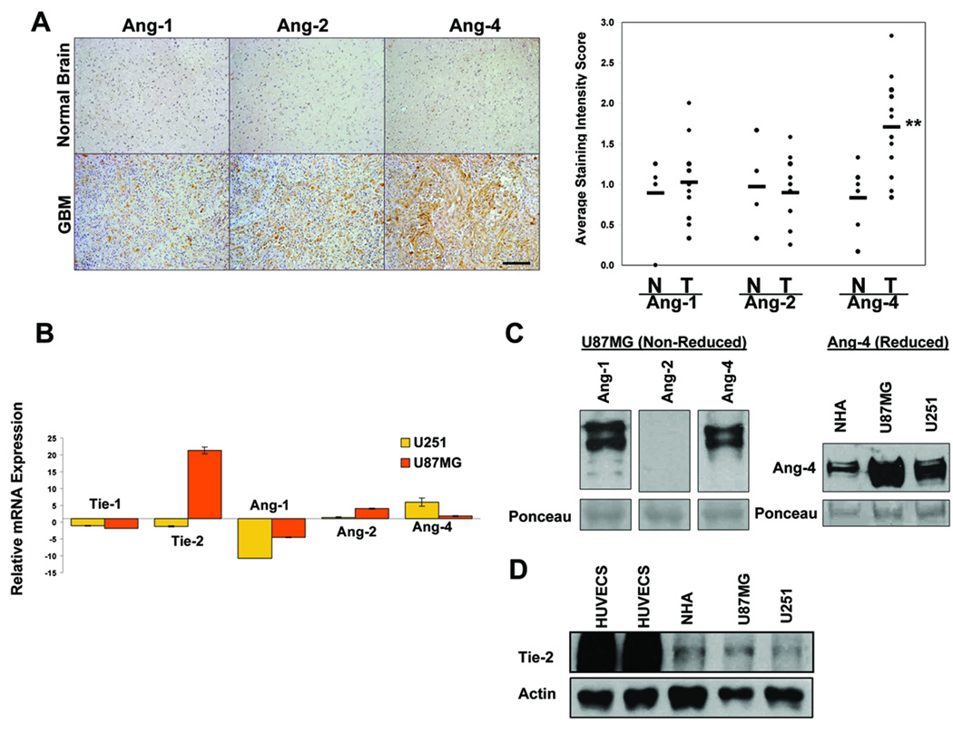
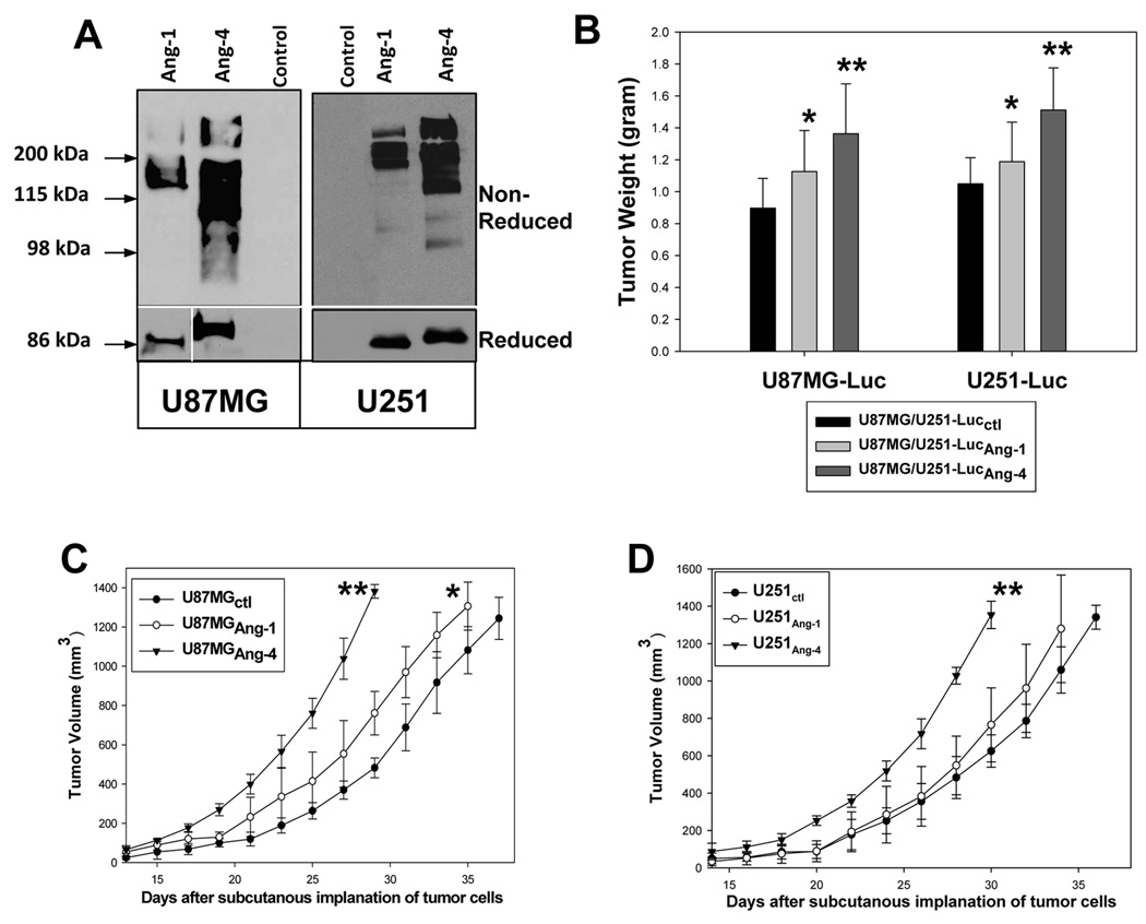
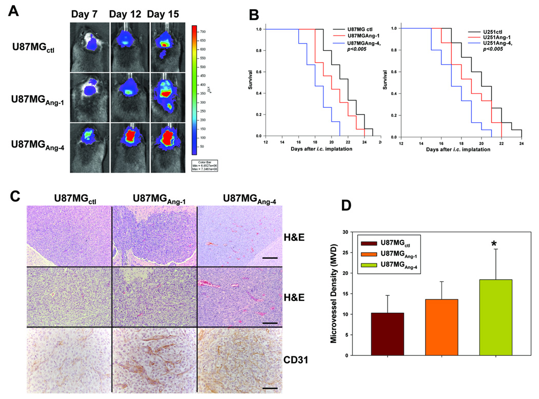
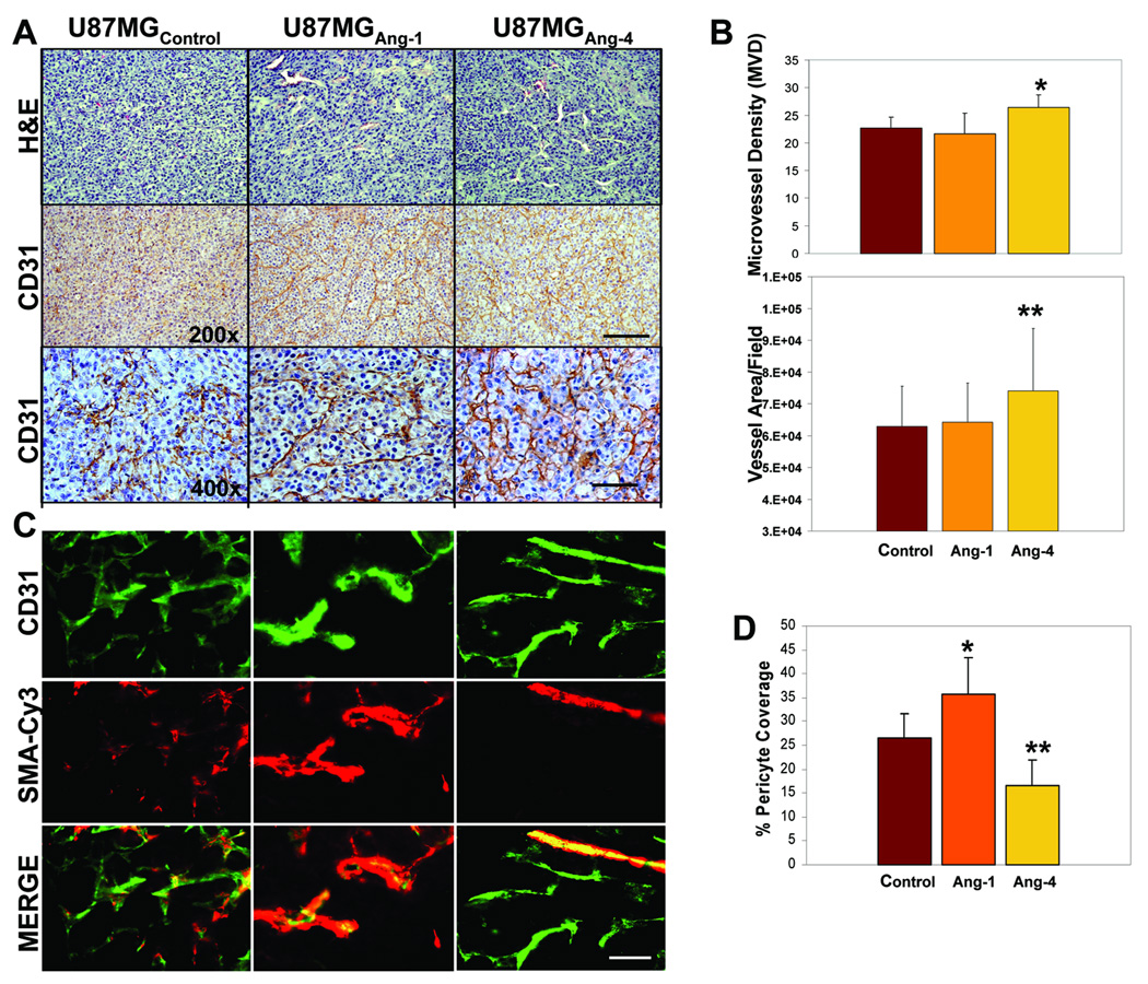
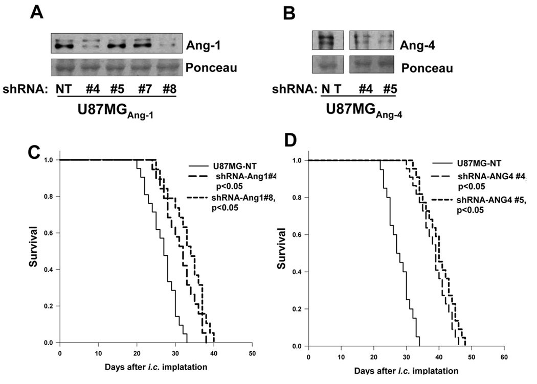
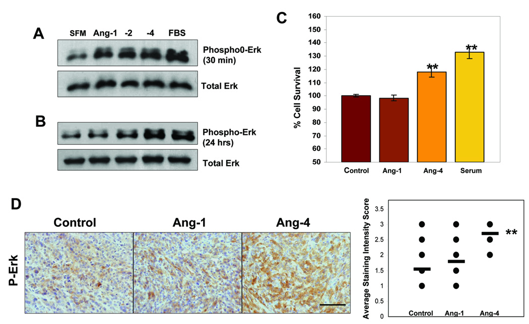
Glioblastoma multiforme (GBM) is a highly invasive and vascularized aggressive brain tumor. Less than 10% of GBM patients survive >5 years after diagnosis. Angiogenesis plays an important role in GBM growth, and antiangiogenesis-based therapies have shown clinical efficacy for GBM patients. Unfortunately, therapeutic resistance often develops in these patients, suggesting that GBM cells are capable of switching their dependency on one proangiogenic signaling pathway to an alternative one. Therefore, it is important to identify novel angiogenic factors that play essential roles in tumor angiogenesis and GBM progression. Angiopoietins (Ang-1, Ang-2, and Ang-4) are the ligands of the Tie-2 receptor tyrosine kinase (RTK). The roles of Ang-1 and Ang-2 in tumor angiogenesis have been established. However, little is known about how Ang-4 affects tumor angiogenesis and GBM progression and the mechanism underlying its effects. In our current study, we establish that Ang-4 is upregulated in human GBM tissues and cells. We show that, like endothelial cells, human GBM cells express Tie-2 RTK. We first establish that Ang-4 promotes in vivo growth of human GBM cells by promoting tumor angiogenesis and directly activating extracellular signal-regulated kinase 1/2 (Erk1/2) in GBM cells. Our results establish the novel effects of Ang-4 on tumor angiogenesis and GBM progression and suggest that this pro-GBM effect of Ang-4 is mediated by promoting tumor angiogenesis and activating Erk1/2 kinase in GBM cells. Together, our results suggest that the Ang-4-Tie-2 functional axis is an attractive therapeutic target for GBM.


Figures

Figure 1
Figure 1. Expression levels of angiopoietins and Tie-2 in GBM tissues and cells
A, Angiopoietin expression was assessed by immunohistochemistry (IHC) using anti-human Ang-1, Ang-2, or Ang-4 antibodies (Santa Cruz). Left panels show representative pictures of 14 GBM samples and 6 normal human brain samples. Bar, 200µm. Right panel shows the plots of intensity scores of the IHC results as determined following the description in Materials and Methods. N = Normal, T = Tumor. B. Real-time qPCR assessing relative transcript levels of human Ang-1, Ang-2, Ang-4, Tie-1, and Tie-2 in U87MG and U251 GBM cells compared to normal human astrocytes (NHAs). C. Endogenous Ang-1, Ang-2, and Ang-4 secreted by U87MG cells (left panel) and endogenous Ang-4 produced by NHAs, U251, and U87MG (right panel) were detected by western blotting with anti-Ang-1 (R&D), Ang-2 (Santa Cruz), or Ang-4 (R&D) antibodies. The SDS-PAGEs were run either under non-reducing conditions (left panel) or reducing conditions (right panel). 160µg of proteins from concentrated serum-free cell culture media was loaded in each lane. The intensities of ~50kDa ponceau-stained bands on the transferred membranes were used as the controls for protein loading and transferring efficiency. D. Expression of endogenous Tie-2 RTK was determined by Western blotting using anti-Tie-2 antibody (Santa Cruz) and the cell lysates derived from human umbilical vein endothelial cells (HUVECs), NHAs, and U87MG and U251 GBM cells (upper panel). 25 µg of total protein was loaded in each lane. Actin was used as a control for protein loading (lower panel).
Figure 2
Figure 2. Ang-4 promotes subcutaneous growth of the GBM cells
A, Establishment of U87MG-Luc and U251-Luc GBM cells expressing v5 epitope tagged Ang-1 (Ang-1v5) or Ang-4 (Ang-4v5). Secreted Ang-1v5 and Ang-4v5 was detected by anti-v5 mAb (Invitrogen) under non-reducing (upper panel) and reducing conditions (bottom panel). B, The effects of Ang-1 and Ang-4 on subcutaneous growth of U87MG and U251 cells were assessed by tumor weight 5 weeks after tumor cell implantation. Six mice were used to implant each type of transduced GBM cells. C–D, Growth rates of the subcutaneous tumors derived from U87MG-Luc (C) and U251-Luc (D) cells expressing Ang-1 or Ang-4 or transduced with empty expression vector. The growth rates are expressed as the mean of tumor volumes (mm3) +/− SD. Six mice were used for each type of transduced GBM cells.
Figure 3
Figure 3. Ang-4 promotes intracranial growth of U87MG-Luc and U251-Luc gliomas
A, Progression of intracranial GBMs was monitored through bio-luminescence imaging at 7, 12, and 15 days after the intracranial injection of the transduced U87MG cells. The images were obtained 12min after injection of D-luciferin using the same intensity scale. B, Survival rates of mice following intracranial injections of the transduced U87MG-Luc (left panel) and U251-Luc (right panel) cells are shown. 15 mice were used for each type of the transduced GBM cells. C, Histologic (H&E) and immunological (anti-CD31) analyses of the GBM sections derived from U87MG-Lucctl, U87MG-LucAng-1, and U87MG-LucAng-4 cells were performed. Bar: 100µm in the upper panels, 50µm in the middle panels, and 25µm in the bottom panels. D, Microvessel density (MVD) in each type of intracranial gliomas was determined by counting the CD31-positive blood vessels in 12 randomly selected 400X microscopic fields within 4 vascular hot-spots.
Figure 4
Figure 4. Ang-4 promotes GBM progression by stimulating tumor angiogenesis
A, Representative H&E and anti-CD31 immunoreactivity pictures of the GBM sections derived from subcutaneous tumors of U87MG-LucAng-1, U87MG-LucAng-4, or U87MG-Lucctl. Bar: 50µm in the upper and middle panels and 25µm in the bottom panels. B, Assessment of tumor angiogenesis: in the top panel, to determine microvessel density (MVD), CD31+ blood vessels within 5 randomly selected 400x microscopic fields in each of 4 vascular hot spots of each type glioma were counted. In the bottom panel, to assess blood vessel area per microscopic field, the perimeter of CD31+ blood vessels from 4 randomly selected 400x microscopic fields in each of 3 vascular hot-spots of each type glioma were outlined and the area was calculated using Image-Pro Plus software. C, The representative pictures of the GBM sections derived from subcutaneous gliomas of U87MG-LucAng-1, U87MG-LucAng-4, or U87MG-Lucctl that show CD31+ ECs and Cy3-smooth muscle actin (SMA)+ peri-vascular cells. Merged pictures of the top and middles panels are shown in the bottom panels. Bar: 20µm. D, Peri-vascular pericyte coverage was determined by assessing numbers of CD 31 positive blood vessels that are covered by Cy3-SMA-positive pericytes in 10 randomly selected 400x microscopic fields of each glioma type. A blood vessel was counted as positive for pericyte coverage when at least 50% of a CD31-positve vessel is covered by SMA-positive pericytes.
Figure 5
Figure 5. Knocking down expression of Ang-4 and to a less extent of Ang-1 inhibits GBM growth in vivo
A–B, Western blots, using anti-Ang-1 (Santa Cruz) or Ang-4 (R&D Systems) antibody, were performed to assess the effectiveness of Ang-1 (A) or Ang-4 (B) knockdown by a panel of TRC-shRNAs (#4, #5, #7, and #8) against human Ang-1 and two TRC-shRNAs (#4 and #5) against human Ang-4. A non-targeting (NT) TRC-shRNA was used as a negative control. 120µg of proteins in concentrated serum free culture supernatants was loaded in each lane. The intensities of ~50kDa ponceau-stained bands were used as the controls for protein loading and transferring efficiency (lower panels). C–D, Survival rates of mice following intracranial injections of the transduced U87MG-Luc with Ang-1 knockdown or NT shRNA (C) or with Ang-4 knockdown or NT shRNA (D). 15 mice were used for each type of transduced GBM cells.
Figure 6
Figure 6. Ang-4 enhances GBM cell viability and actives Erk1/2 kinase in vitro and in vivo
A–B, Serum-starved U87MG cells were supplied with serum free medium (SFM), 10%FBS containing medium, or SFM containing 200ng/ml of Ang-1 or Ang-4 for 30min (A) and 24 hours (B). The cells were lysed and proteins were analyzed by Western blotting with anti-phospho-Erk1/2 (upper panels) or with anti-Erk1/2 (bottom panels) antibodies. C, Cell viability assays were performed. U87MG cells were plated at 2 × 10⁴/well of 96-well plates in triplicates and allowed to grow overnight. The cells were switched to SFM and cultured for an additional 48 hrs and then treated with 200ng/ml of Ang-1 or Ang-4, 10%FBS, or SFM for another 24 hrs before cell viability was measured using the Cell Titer-Glo Luminescent Cell Viability Assay kit (Promega) following the manufacturer’s instructions. D, Levels of phosphorylated Erk1/2 in the GBM sections derived from subcutaneous tumors of U87MG-LucAng-1, U87MG-LucAng-4, or U87MG-Lucctl were detected using anti-phopho-Erk1/2 antibody (Santa Cruz). The immunoreactivity intensity was scored in ten randomly selected 400x microscopic filed using a 0–3 scoring system described in Materials and Methods.

Similar articles

Cited by

References

    1. Norden AD, Drappatz J, Wen PY. Novel anti-angiogenic therapies for malignant gliomas. Lancet Neurol. 2008;7:1152–1160. - PubMed
    1. Norden AD, Drappatz J, Wen PY. Antiangiogenic therapy in malignant gliomas. Curr Opin Oncol. 2008;20:652–661. - PubMed
    1. Norden AD, Young GS, Setayesh K, et al. Bevacizumab for recurrent malignant gliomas: efficacy, toxicity, and patterns of recurrence. Neurology. 2008;70:779–787. - PubMed
    1. Masabumi S. Brain angiogenesis in developmental and pathological processes: therapeutic aspects of vascular endothelial growth factor. FEBS Journal. 2009;276:4636–4643. - PubMed
    1. Woo-Young K, Ho-Young L. Brain angiogenesis in developmental and pathological processes: mechanism and therapeutic intervention in brain tumors. FEBS Journal. 2009;276:4653–4664. - PMC - PubMed

Publication types

MeSH terms