. 2010 Sep 15;70(18):7042-52.
doi: 10.1158/0008-5472.CAN-10-0522. Epub 2010 Sep 7.

The sympathetic nervous system induces a metastatic switch in primary breast cancer

Affiliations

The sympathetic nervous system induces a metastatic switch in primary breast cancer

Erica K Sloan et al. Cancer Res. .

Abstract

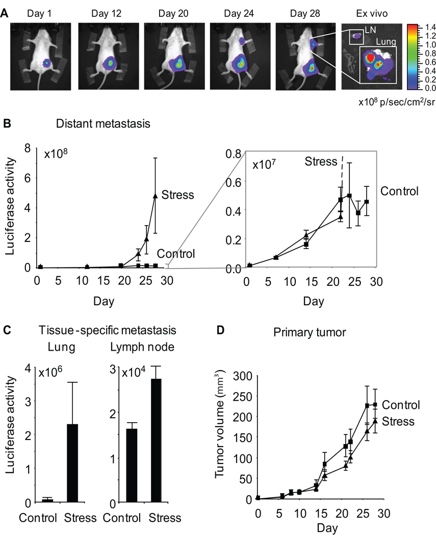
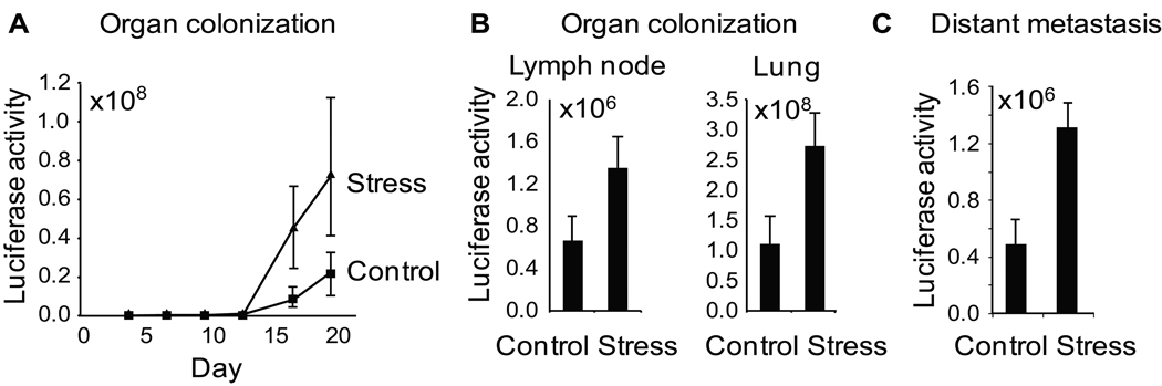
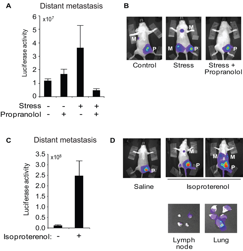
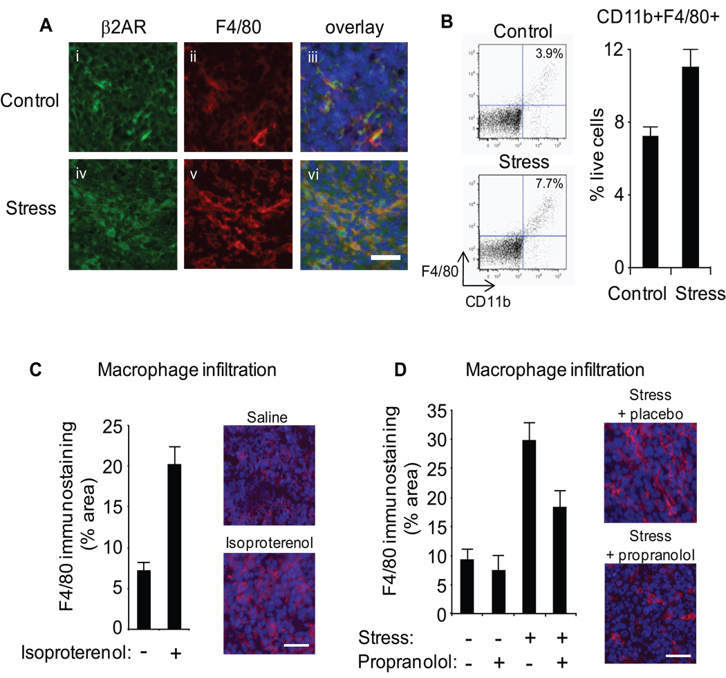
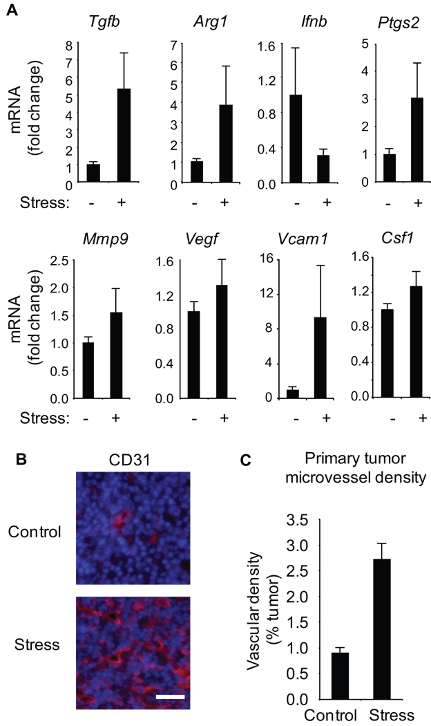
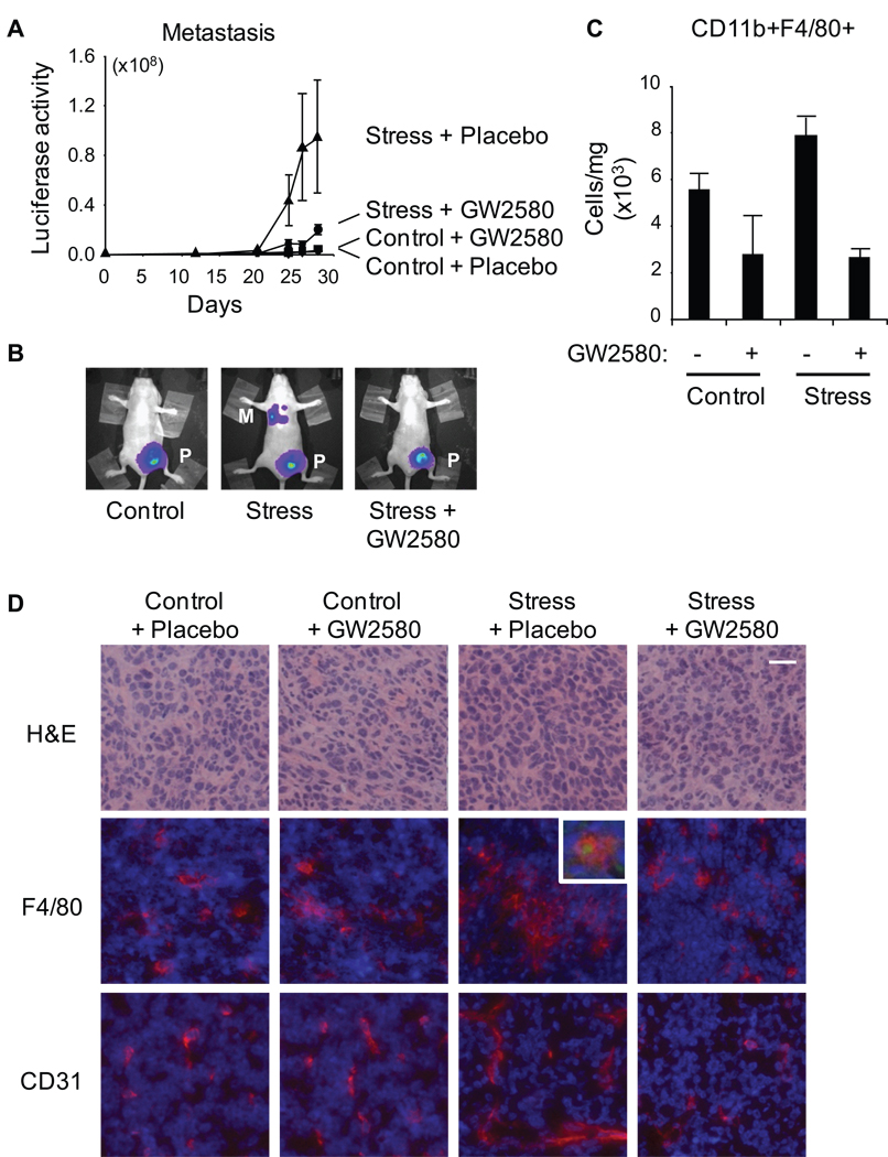
Metastasis to distant tissues is the chief driver of breast cancer-related mortality, but little is known about the systemic physiologic dynamics that regulate this process. To investigate the role of neuroendocrine activation in cancer progression, we used in vivo bioluminescence imaging to track the development of metastasis in an orthotopic mouse model of breast cancer. Stress-induced neuroendocrine activation had a negligible effect on growth of the primary tumor but induced a 30-fold increase in metastasis to distant tissues including the lymph nodes and lung. These effects were mediated by β-adrenergic signaling, which increased the infiltration of CD11b(+)F4/80(+) macrophages into primary tumor parenchyma and thereby induced a prometastatic gene expression signature accompanied by indications of M2 macrophage differentiation. Pharmacologic activation of β-adrenergic signaling induced similar effects, and treatment of stressed animals with the β-antagonist propranolol reversed the stress-induced macrophage infiltration and inhibited tumor spread to distant tissues. The effects of stress on distant metastasis were also inhibited by in vivo macrophage suppression using the CSF-1 receptor kinase inhibitor GW2580. These findings identify activation of the sympathetic nervous system as a novel neural regulator of breast cancer metastasis and suggest new strategies for antimetastatic therapies that target the β-adrenergic induction of prometastatic gene expression in primary breast cancers.


Figures

Figure 1
Figure 1. Effect of chronic stress on breast cancer metastasis to distant tissues
A. Metastasis was quantified by periodic imaging of tumor-specific bioluminescence signal in chest region in Balb/c mice and (B) quantified over multiple mice (5 per group). Inset shows increased resolution of the control condition. Luciferase activity: photons/sec. C. Tissue-specific metastasis was quantified by ex vivo bioluminescent imaging of tumor masses in lung and lymph node (axillary and brachial). D. Primary tumor volume was derived from 2-dimensional caliper measurements. Data: Mean ± S.E.
Figure 2
Figure 2. The effect of stress on colonization of metastatic target tissues
A. Organ colonization by 1.0 × 10⁵ i.v-injected 66cl4 tumor cells was quantified by repeated imaging of tumor-specific bioluminescence signal in chest region. B. Tissue specific colonization was quantified by ex vivo imaging of lymph node and lung on day 27 after intravenous inoculation of tumor cells. C. Distant metastasis was quantified by bioluminescence imaging at day 41 following surgical removal of primary tumors on day 14. Stress was commenced only after surgical removal of primary tumors. Data: Mean ± S.E.
Figure 3
Figure 3. Role of sympathetic nervous system in stress-induced metastasis
A. Distant metastasis was quantified in the lung and lymph node region by in vivo imaging at day 28 following tumor cell inoculation into stressed vs control mice treated with propranolol or placebo. B. Representative images of mice in control (+ placebo), stress (+ placebo) and stress + beta-blocker groups. C. Distant metastasis was quantified 28 days after primary tumor inoculation in mice treated with saline vs isoproterenol. D. Representative in vivo images of saline- and isoproterenol-treated mice and (E) ex vivo lung and lymph node showing tumor-specific bioluminescent signal (× 106 p/sec/cm2/sr). P: primary tumor, M: distant metastasis.
Figure 4
Figure 4. Role of macrophage infiltration
A. Mammary tumor cryosections from control and stressed mice were immunostained with anti-β2AR (green) (i, iv) and anti-F4/80 (red) (ii, v), and nuclear counterstained with Hoechst 33324 (blue) (iii, vi). B. CD11b+F4/80+ cells were quantified by flow cytometry in disaggregated 66cl4 primary tumors harvested 28 days after inoculation. C,D. Macrophage infiltration of mammary tumors was visualized by immunostaining with anti-F4/80 (red) and nuclei counterstained (blue) in tumors harvested at day 28 from saline- or isoproterenol-treated mice (C) or stressed mice treated with placebo vs. propranolol (D), and quantified across sections from multiple mice (n = 5/gp). Scale bars: 30µm.
Figure 5
Figure 5. Effect of stress on pro-metastatic gene expression and primary tumor vascularization
A. Gene expression was quantified by RT-PCR in mammary tumors from control vs. stressed mice (n = 5/gp). B. Mammary tumor cryosections were immunostained with anti-CD31 antibody (red) and nuclei counterstained (blue). Scale bar: 30 µm. C. CD31+ blood vessel density was quantified in 5 sections from multiple tumors (n = 5/gp).
Figure 6
Figure 6. Effect of macrophage inhibition on stress-enhanced metastasis
A. Metastasis was quantified using live in vivo imaging in control vs stressed mice treated with the CSF1 receptor inhibitor GW2580 or placebo. (NB: Control + placebo and control + GW2580 curves are indistinguishable.) B. Representative images of mice in control (+ placebo), stress (+ placebo) and stress + GW2580 conditions. C. Frequency of CD11b+F4/80+ cells in 66cl4 primary tumors from control vs stressed mice treated with GW2580 or placebo. D. Mammary tumor cryosections were stained with hematoxylin and eosin to show general morphology or immunostained with anti-F4/80 (red, middle panels) or anti-CD31 (red, lower panels) and nuclei counterstained (blue). Scale bar: 20µm. Inset shows representative F4/80 macrophages positive for β2AR (green). P: primary tumor, M: distant metastasis.

References

    1. Antoni MH, Lutgendorf SK, Cole SW, Dhabhar FS, Sephton SE, McDonald PG, et al. The influence of bio-behavioural factors on tumour biology: pathways and mechanisms. Nat Rev Cancer. 2006;6:240–248. - PMC - PubMed
    1. Thaker PH, Han LY, Kamat AA, Arevalo JM, Takahashi R, Lu C, et al. Chronic stress promotes tumor growth and angiogenesis in a mouse model of ovarian carcinoma. Nat Med. 2006;12:939–944. - PubMed
    1. Sloan EK, Capitanio JP, Tarara RP, Mendoza SP, Mason WA, Cole SW. Social stress enhances sympathetic innervation of primate lymph nodes: mechanisms and implications for viral pathogenesis. J Neurosci. 2007;27:8857–8865. - PMC - PubMed
    1. Tang Y, Shankar R, Gamelli R, Jones S. Dynamic norepinephrine alterations in bone marrow: evidence of functional innervation. J Neuroimmunol. 1999;96:182–189. - PubMed
    1. Cakir Y, Plummer HK, 3rd, Tithof PK, Schuller HM. Beta-adrenergic and arachidonic acid-mediated growth regulation of human breast cancer cell lines. Int J Oncol. 2002;21:153–157. - PubMed

Publication types