. 2010 Sep 15;70(18):7053-62.
doi: 10.1158/0008-5472.CAN-10-0271. Epub 2010 Sep 7.

Quantitative imaging of lymphatic function with liposomal indocyanine green

Affiliations

Quantitative imaging of lymphatic function with liposomal indocyanine green

Steven T Proulx et al. Cancer Res. .

Abstract

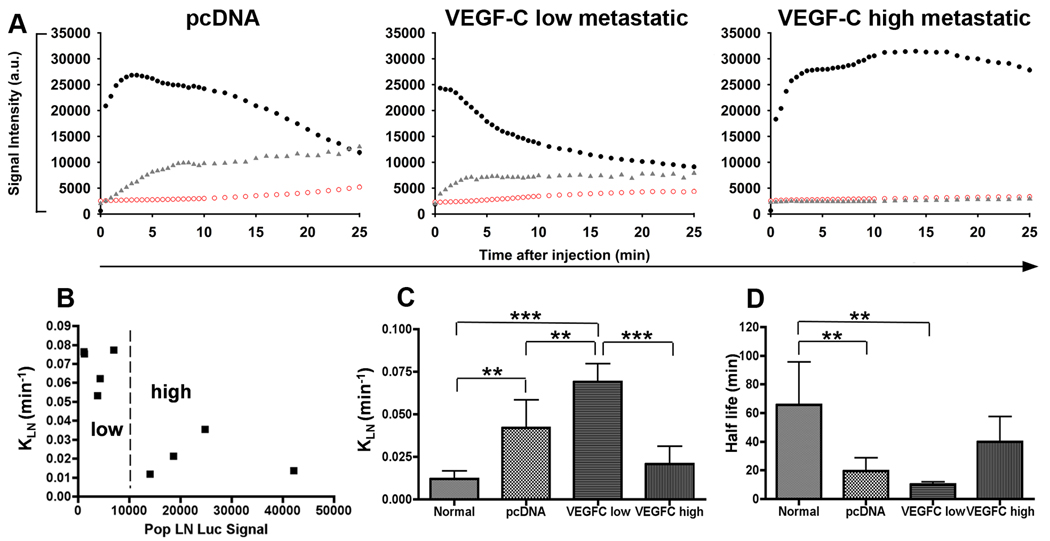
Lymphatic vessels play a major role in cancer progression and in postsurgical lymphedema, and several new therapeutic approaches targeting lymphatics are currently being developed. Thus, there is a critical need for quantitative imaging methods to measure lymphatic flow. Indocyanine green (ICG) has been used for optical imaging of the lymphatic system, but it is unstable in solution and may rapidly enter venous capillaries after local injection. We developed a novel liposomal formulation of ICG (LP-ICG), resulting in vastly improved stability in solution and an increased fluorescence signal with a shift toward longer wavelength absorption and emission. When injected intradermally to mice, LP-ICG was specifically taken up by lymphatic vessels and allowed improved visualization of deep lymph nodes. In a genetic mouse model of lymphatic dysfunction, injection of LP-ICG showed no enhancement of draining lymph nodes and slower clearance from the injection site. In mice bearing B16 luciferase-expressing melanomas expressing vascular endothelial growth factor-C (VEGF-C), sequential near-IR imaging of intradermally injected LP-ICG enabled quantification of lymphatic flow. Increased flow through draining lymph nodes was observed in mice bearing VEGF-C-expressing tumors without metastases, whereas a decreased flow pattern was seen in mice with a higher lymph node tumor burden. This new method will likely facilitate quantitative studies of lymphatic function in preclinical investigations and may also have potential for imaging of lymphedema or improved sentinel lymph detection in cancer.


Figures

Figure 1
Figure 1. Spectral properties of LP-ICG
Typical normalized absorption (A) and fluorescence (B) spectra of LP-ICG (C) with respect to free ICG. Solid line: ICG in 5% glucose; dashed line: LP-ICG. The spectra are normalized for the maximum absorbance (λmax = 780 nm) and the maximum fluorescence emission (λmax = 810 nm) of free ICG. The stability of the spectral properties of LP-ICG over time is compared to that of ICG in buffer. The fluorescence maximum emission over time (D) is normalized for the initial maximum emission (ICG λem = 810 nm; LP-ICG λem = 831 nm). Filled circles: ICG; empty circles: LP-ICG.
Figure 2
Figure 2. Dynamics of ICG signal after intradermal injection in normal C57BL/6 albino mice
Time series of images from representative animals before and after intradermal injection of the left rear paw with ICG (A) and LP-ICG (B). Images were collected with 4-s exposure time in both animals. Enhancement of popliteal lymph node and liver are seen in (A) and (B), while the medial iliac lymph node shows also signal enhancement in (B). Signal intensity of enhanced tissues is plotted versus time in ICG (C) and LP-ICG (D) injected mice. Black circles: popliteal lymph node; red squares: liver; grey triangles: medial iliac lymph node.
Figure 3
Figure 3. NIRF imaging of Chy mice after intradermal injection
Time series of images (exposure time 6 s) after intradermal injection of LP-ICG in a representative 5 month old male Chy mouse (A, left) and wild type littermate (A, right). Images of injection site (exposure time 0.1 s) taken 10 min, 4 h, and 24 h after injection into foot of a representative Chy mouse (B, left) and wild type littermate (B, right). White arrowhead at 24 h in (B, left) shows interstitial diffusion pattern from foot in Chy mouse. In C, scatter plots of signal intensity measurements of injected feet at 10 min, 4 h, and 24 h. * p < 0.05
Figure 4
Figure 4. Bioluminescent imaging of popliteal lymph node metastases in human VEGF-C overexpressing B16-luc2 melanoma tumor bearing mice
(A) Tumor volumes of VEGF-C expressing and control cells at 21 days. Quantification of in vivo bioluminescent signals from 9 VEGF-C and 8 pcDNA tumor bearing mice (B). Bioluminescent images of popliteal lymph node region revealing lymph node metastasis in representative VEGF-C tumor bearing mouse (C) and lack of signal in pcDNA tumor bearing mouse (D). Insets: Ex vivo signal of dissected popliteal lymph nodes from VEGF-C (C) or pcDNA (D) tumor bearing mice. * p < 0.05
Figure 5
Figure 5. Dynamic NIRF imaging of ICG liposome injected B16-luc2 tumor bearing mice
Representative figures of lymphatic drainage patterns of LP-ICG after intradermal injection (A) from pcDNA, VEGF-C low metastatic, and VEGF-C high metastatic mice. Black circles: popliteal lymph node; red squares: liver; grey triangles: medial iliac lymph node. Correlation plot (B) showing KLN rates through popliteal lymph node versus popliteal lymph node luciferase signals. Dashed line represents threshold between low metastatic and high metastatic VEGF-C tumor bearing mice. KLN rates (C) and half life measurements (D) of normal, pcDNA tumor, VEGF-C low metastatic and VEGF-C high metastatic groups. * p < 0.05, ** p < 0.01, *** p < 0.001.

References

    1. Karpanen T, Alitalo K. Molecular biology and pathology of lymphangiogenesis. Annu Rev Pathol. 2008;3:367–397. - PubMed
    1. Jurisic G, Detmar M. Lymphatic endothelium in health and disease. Cell Tissue Res. 2009;335:97–108. - PubMed
    1. Mattila MM-T, Ruohola JK, Karpanen T, Jackson DG, Alitalo K, Härkönen PL. VEGF-C induced lymphangiogenesis is associated with lymph node metastasis in orthotopic MCF-7 tumors. Int J Cancer. 2002;98:946–951. - PubMed
    1. Mumprecht V, Detmar M. Lymphangiogenesis and cancer metastasis. J Cell Mol Med. 2009;13:1405–1416. - PMC - PubMed
    1. Skobe M, Hawighorst T, Jackson DG, et al. Induction of tumor lymphangiogenesis by VEGF-C promotes breast cancer metastasis. Nat Med. 2001;7:192–198. - PubMed

Publication types

MeSH terms