. 2010 Nov;78(11):4454-66.
doi: 10.1128/IAI.00840-10. Epub 2010 Sep 7.

Mannheimia haemolytica and its leukotoxin cause neutrophil extracellular trap formation by bovine neutrophils

Affiliations

Mannheimia haemolytica and its leukotoxin cause neutrophil extracellular trap formation by bovine neutrophils

Nicole A Aulik et al. Infect Immun. 2010 Nov.

Abstract

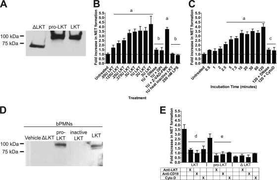
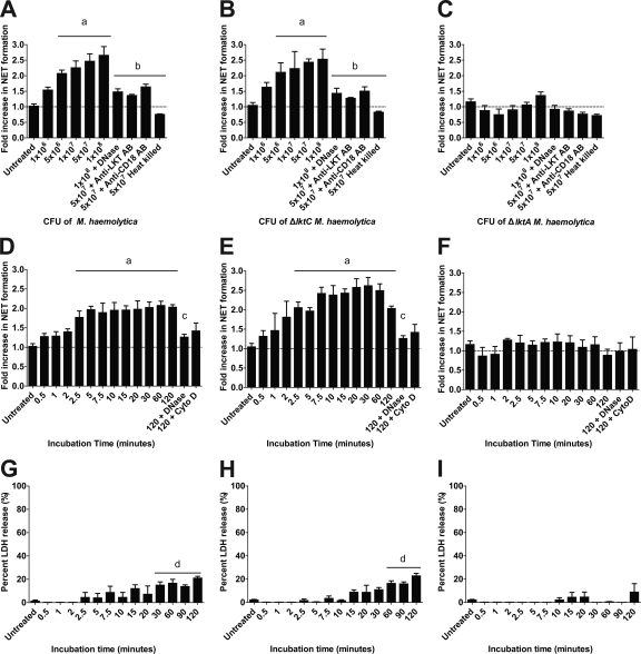
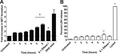
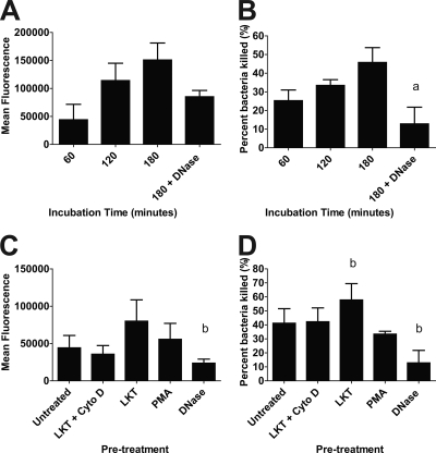
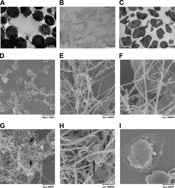
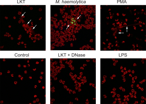
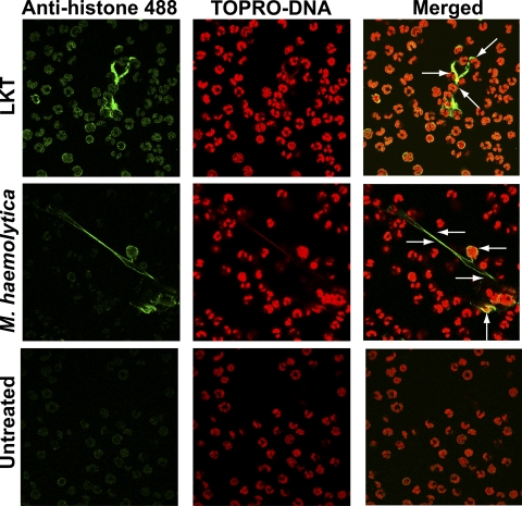
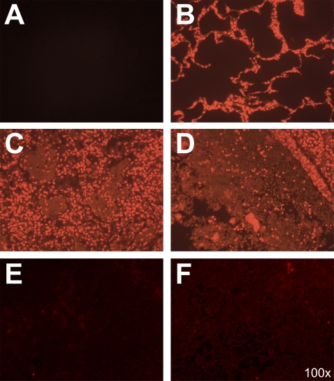
Mannheimia haemolytica is an important member of the bovine respiratory disease complex, which is characterized by abundant neutrophil infiltration into the alveoli and fibrin deposition. Recently several authors have reported that human neutrophils release neutrophil extracellular traps (NETs), which are protein-studded DNA matrices capable of trapping and killing pathogens. Here, we demonstrate that the leukotoxin (LKT) of M. haemolytica causes NET formation by bovine neutrophils in a CD18-dependent manner. Using an unacylated, noncytotoxic pro-LKT produced by an ΔlktC mutant of M. haemolytica, we show that binding of unacylated pro-LKT stimulates NET formation despite a lack of cytotoxicity. Inhibition of LKT binding to the CD18 chain of lymphocyte function-associated antigen 1 (LFA-1) on bovine neutrophils reduced NET formation in response to LKT or M. haemolytica cells. Further investigation revealed that NETs formed in response to M. haemolytica are capable of trapping and killing a portion of the bacterial cells. NET formation was confirmed by confocal microscopy and by scanning and transmission electron microscopy. Prior exposure of bovine neutrophils to LKT enhanced subsequent trapping and killing of M. haemolytica cells in bovine NETs. Understanding NET formation in response to M. haemolytica and its LKT provides a new perspective on how neutrophils contribute to the pathogenesis of bovine respiratory disease.


Figures

FIG. 1.
FIG. 1.
LKT and pro-LKT cause NET formation by bovine neutrophils. (A) Purified native LKT, pro-LKT, and ΔLKT were separated by SDS-PAGE, blotted to PVDF, and probed using an anti-LKT antibody to estimate the molecular masses of the toxins. (B and C) bPMNs (106) were incubated with various amounts of LKT for 120 min (B) or were incubated with 1 U of LKT for various times (C). As a control, 106 bPMNs were preincubated with 180 U of DNase I, 10 μg/ml cytochalasin D (Cyto D), 10 μM DPI, or 200 μM Z-VAD-FMK (pan-caspase inhibitor) before LKT was added. (D) bPMNs (106) were incubated with 1 U LKT, heat-inactivated LKT, pro-LKT, or ΔLKT for 120 min. Cells were then washed, and lysates were prepared. Proteins were separated by SDS-PAGE, blotted to PVDF, and probed using an anti-LKT antibody. Untreated control cells were preincubated with the vehicle RPMI 1640. (E) bPMNs (106) were preincubated for 2 h with 1 U LKT, pro-LKT, or ΔLKT. Some bPMNs were incubated with an anti-CD18 antibody or 10 μg/ml cytochalasin D, or toxins were incubated with an anti-LKT antibody for 30 min prior to addition to bPMNs. Extracellular DNA was quantified by the addition of a 1:200 dilution of PicoGreen, and fluorescence was quantified using an automated plate reader. Data represent the means ± standard errors of the means of 5 independent experiments. a, P < 0.05 compared to untreated bPMNs; b, P < 0.05 compared to bPMNs treated with 1 U LKT; c, P < 0.05 compared to bPMNs treated with LKT for 120 min; d, P < 0.05 compared to bPMNs incubated with LKT; e, P < 0.05 compared to bPMNs incubated with pro-LKT.
FIG. 2.
FIG. 2.
Wild-type M. haemolytica and ΔlktC M. haemolytica cells cause NET formation by bovine neutrophils. (A to C) bPMNs (106) were incubated for 120 min with various numbers of M. haemolytica (A), ΔlktC M. haemolytica (B), or ΔlktA M. haemolytica (C) cells. In some experiments, 180 U of DNase I or an anti-CD18 antibody was preincubated with the bPMNs, or bacteria were preincubated with an anti-LKT antibody, for 30 min prior to addition to bPMNs. (D to F) bPMNs (106) were incubated for various times with 107 cells of M. haemolytica (D), ΔlktC M. haemolytica (E), or ΔlktA M. haemolytica (F). In some experiments, 180 U of DNase I or 10 μg/ml cytochalasin D (Cyto D) was preincubated with the bPMNs for 30 min. Extracellular DNA was quantified as described previously. (G to I) bPMNs (106) were incubated with 107 CFU of M. haemolytica (G), ΔlktC M. haemolytica (H), or ΔlktA M. haemolytica (I), and LDH release was quantified using the CytoTox 96 nonradioactive cytotoxicity assay as described by the manufacturer. Data represent the means ± standard errors of the means of 5 independent experiments. a, P < 0.05 compared to untreated bPMNs; b, P < 0.05 compared to bPMNs incubated with 5 × 10⁷ bacteria; c, P < 0.05 compared to bPMNs incubated for 120 min with bacterial cells alone; d, P < 0.05 compared to LDH release by untreated bPMNs.
FIG. 3.
FIG. 3.
Repeated exposure to M. haemolytica results in increased NET formation by bovine neutrophils. M. haemolytica cells (107) were added to 106 bPMNs every hour for 1 to 6 h and NET formation (A) and LDH release (B) were measured at 6 h. DNase I (180 U) was also added to one group of cells that was treated hourly for 6 h. Total DNA was determined by lysing 106 bPMNs, and extracellular DNA was quantified as described previously. Data represent the means ± standard errors of the means of 5 independent experiments. a, P < 0.05 compared to bPMNs treated for 1 h; b, P < 0.05 compared to bPMNs treated for 6 h; c, P < 0.05 compared to untreated bPMNs.
FIG. 4.
FIG. 4.
Preincubating bovine neutrophils with LKT increases the trapping and killing of M. haemolytica within NETs. (A and B) bPMNs (106) were incubated with 107 fluorescein-labeled M. haemolytica (A) or unlabeled M. haemolytica (B) cells for 60 to 180 min. (C and D) Similarly, treated bPMNs (LKT plus Cyto D, LKT, PMA, or DNase) or untreated bPMNs were incubated with 107 fluorescein-labeled M. haemolytica cells (C) or unlabeled M. haemolytica cells (D) for 180 min. In panels C and D, 106 bPMNs were incubated with (i) 0.25 U LKT for 30 min followed by 10 μg/ml cytochalasin D (LKT + Cyto D), (ii) 0.25 U LKT, (iii) 100 nM PMA for 30 min at 37°C, or (iv) 180 U DNase I for the duration of the experiment. Fluorescence (A and C) was measured using an automated plate reader. Bacterial survival (B and D) was estimated by plating serial dilutions of lysates on TSA plus 5% sheep RBC plates. Data represent the means ± standard errors of the means of 5 independent experiments. a, P < 0.05 compared to bPMNs treated for 180 min; b, P < 0.05 compared to untreated bPMNs.
FIG. 5.
FIG. 5.
Transmission and scanning electron photomicrographs of NETs formed by bovine neutrophils in response to M. haemolytica LKT. LKT (0.5 U) or 5 × 10⁷ M. haemolytica cells were incubated with 3 × 10⁶ bPMNs for 60 min at 37°C. Cells were washed, fixed, and processed for TEM (A to C) or SEM (D to I) as described in Materials and Methods. Transmission electron photomicrographs were taken at a magnification of ×3,400 (bar, 5 μm) (A and C) or ×17,000 (bar, 200 nm) (B). (A) Large weblike structures were released from LKT-treated bPMNs. (B) Enlargement of panel A, to illustrate individual fibrils. (C) Control cells incubated in RPMI medium do not exhibit fibrils. (D to I) Scanning electron photomicrographs. (D) Micrograph demonstrates a large network of extracellular DNA strands released in response to LKT. (E) Higher magnification of panel D, illustrating bPMNs trapped within the mesh. (F) Higher magnification of panel E, illustrating in greater detail the ultrastructure of individual NET fibrils. (G) Complex network of DNA strands in which bPMNs and M. haemolytica cells are visible. (H) Higher magnification of panel G, illustrating more closely M. haemolytica cells trapped within a NET. Arrows indicate trapped M. haemolytica cells in panels G and H. (I) Control bPMNs incubated in RPMI medium do not exhibit fibrils. Photomicrographs are of representative cells from 3 independent experiments.
FIG. 6.
FIG. 6.
Confocal photomicrographs reveal extracellular DNA released from bovine neutrophils in response to LKT, M. haemolytica cells, or PMA. bPMNs (3 × 10⁶) were allowed to attach to glass slides and then were incubated with 0.5 U LKT, 5 × 10⁷ fluorescein-labeled M. haemolytica cells, or 100 nM PMA. Controls include untreated bPMNs (control) and bPMNs incubated with 0.5 U LKT plus 180 U DNase I, or 250 nM LPS, for 60 min. Cells were fixed, permeabilized, stained for DNA using TOPRO, and examined by confocal microscopy. Arrows illustrate representative NETs. Photomicrographs are of representative cells from 3 independent experiments.
FIG. 7.
FIG. 7.
Colocalization of DNA and histones in NETs produced in response to LKT or M. haemolytica. bPMNs (3 × 10⁶) were allowed to attach to glass slides and then incubated with 0.5 U LKT or 5 × 10⁷ M. haemolytica cells for 60 min. Cells were fixed, permeabilized, stained for DNA using TOPRO (red), and probed for histones using an antihistone antibody followed by an anti-mouse antibody labeled with Alexa Fluor 488 (green). Cells were examined by confocal microscopy. Arrows indicate areas of colocalization of signal (yellow-orange) for extracellular DNA and histones. Photomicrographs are of representative cells from 3 independent experiments.
FIG. 8.
FIG. 8.
Extracellular DNA is present in the lungs of an M. haemolytica-infected calf. Tissue sections (10 μm) were obtained from an M. haemolytica-infected calf and a healthy calf, deparaffinized, and incubated with Sytox Orange to stain DNA, or PBS as a control for autofluorescence at 570 nm. (A) Control lung tissue from an M. haemolytica-infected calf that was not stained with Sytox Orange to illustrate the absence of autofluorescence at 570 nm. (B) Lung section from a healthy bovine lung displays normal alveolar structure with no extracellular DNA staining. (C and D) Lung tissue obtained from an M. haemolytica-infected calf that was stained with Sytox Orange to detect DNA. (C) Cellular infiltrates in the alveoli with extracellular DNA present. (D) Consolidation of the lung and extensive extracellular DNA staining. (E and F) Lung tissues from an M. haemolytica-infected calf were incubated for 24 h with 180 U DNase at 4°C, washed, and incubated with Sytox Orange. Fluorescence was reduced compared to that in panels C and D, indicating that the Sytox staining was specific for DNA within the tissue.
FIG. 9.
FIG. 9.
Coincubation with bovine serum causes a reduction in NET formation. bPMNs (106) were incubated with 1 U LKT (A), 107 M. haemolytica cells (B), 107 ΔlktC M. haemolytica cells (C), or 107 ΔlktA M. haemolytica cells (D) for 120 min with increasing amounts of normal adult bovine serum. In some experiments, 180 U of DNase I was also added to the 20% serum group. Extracellular DNA was quantified as described above. Data represent the means ± standard errors of the means of 5 independent experiments. a, P < 0.05 compared to bPMNs incubated without bovine serum.

References

    1. Ackermann, M. R., B. M. DeBey, T. J. Stabel, J. H. Gold, K. B. Register, and J. T. Meehan. 1994. Distribution of anti-CD68 (EBM11) immunoreactivity in formalin-fixed, paraffin-embedded bovine tissues. Vet. Pathol. 31:340-348. - PubMed
    1. Alexander, L. E., H. C. Maisey, A. M. Timmer, S. H. Rooijakkers, R. L. Gallo, M. von Kockritz-Blickwede, and V. Nizet. 2010. M1T1 group A streptococcal pili promote epithelial colonization but diminish systemic virulence through neutrophil extracellular entrapment. J. Mol. Med. 88:371-381. - PMC - PubMed
    1. Ambagala, T. C., A. P. Ambagala, and S. Srikumaran. 1999. The leukotoxin of Pasteurella haemolytica binds to beta(2) integrins on bovine leukocytes. FEMS Microbol. Lett. 179:161-167. - PubMed
    1. Atapattu, D. N., R. M. Albrecht, D. J. McClenahan, and C. J. Czuprynski. 2008. Dynamin-2-dependent targeting of Mannheimia haemolytica leukotoxin to mitochondrial cyclophilin D in bovine lymphoblastoid cells. Infect. Immun. 76:5357-5365. - PMC - PubMed
    1. Atapattu, D. N., and C. J. Czuprynski. 2007. Mannheimia haemolytica leukotoxin binds to lipid rafts in bovine lymphoblastoid cells and is internalized in a dynamin-2- and clathrin-dependent manner. Infect. Immun. 75:4719-4727. - PMC - PubMed

Publication types

MeSH terms

LinkOut - more resources