. 2010 Oct 5;107(40):17339-44.
doi: 10.1073/pnas.1010026107. Epub 2010 Sep 7.

Rigid amphipathic fusion inhibitors, small molecule antiviral compounds against enveloped viruses

Affiliations

Rigid amphipathic fusion inhibitors, small molecule antiviral compounds against enveloped viruses

Mireille R St Vincent et al. Proc Natl Acad Sci U S A. .

Abstract

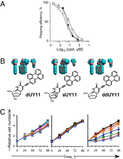
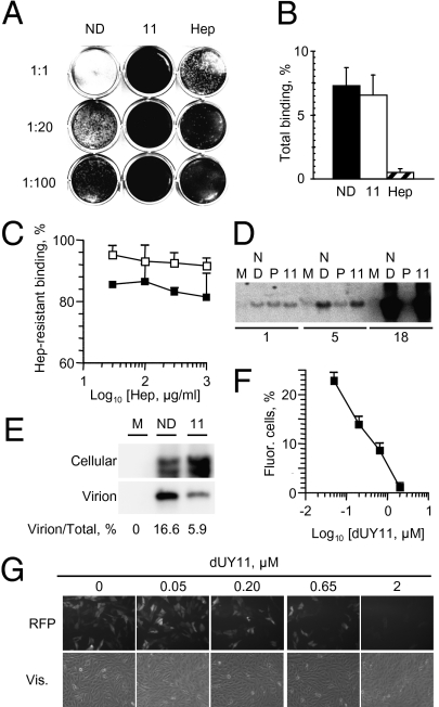
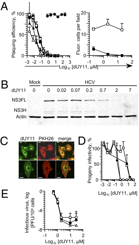
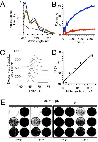
Antiviral drugs targeting viral proteins often result in prompt selection for resistance. Moreover, the number of viral targets is limited. Novel antiviral targets are therefore needed. The unique characteristics of fusion between virion envelopes and cell membranes may provide such targets. Like all fusing bilayers, viral envelopes locally adopt hourglass-shaped stalks during the initial stages of fusion, a process that requires local negative membrane curvature. Unlike cellular vesicles, however, viral envelopes do not redistribute lipids between leaflets, can only use the energy released by virion proteins, and fuse to the extracellular leaflets of cell membranes. Enrichment in phospholipids with hydrophilic heads larger than their hydrophobic tails in the convex outer leaflet of vesicles favors positive curvature, therefore increasing the activation energy barrier for fusion. Such phospholipids can increase the activation barrier beyond the energy provided by virion proteins, thereby inhibiting viral fusion. However, phospholipids are not pharmacologically useful. We show here that a family of synthetic rigid amphiphiles of shape similar to such phospholipids, RAFIs (rigid amphipathic fusion inhibitors), inhibit the infectivity of several otherwise unrelated enveloped viruses, including hepatitis C and HSV-1 and -2 (lowest apparent IC(50) 48 nM), with no cytotoxic or cytostatic effects (selectivity index > 3,000) by inhibiting the increased negative curvature required for the initial stages of fusion.


Conflict of interest statement

Conflict of interest statement: M.R.S., A.V.U., and L.M.S. are co-inventors in a series of patent applications describing these discoveries.

Figures

Fig. 1.
Fig. 1.
Rigid amphipathic nucleoside derivatives inhibit HSV-1 infectivity with no cytotoxic effects. (A) Plaquing efficiency of HSV-1 virions preexposed to increasing concentrations of dUY11 (□), aUY11 (○), or ddUY11(▲) (average ± SD; n ≥ 5). (B) Chemical structures and 3D models, displayed in three orthogonal perspectives, of dUY11, aUY11, and ddUY11. Gray, carbon; teal, hydrogen; pink, oxygen; blue, nitrogen. (C) Vero cell viability in the presence of dUY11, aUY11, or ddUY11. Relative numbers of viable cells plotted against time of treatment with 0 (black squares), 0.05 (sky blue circles), 0.15 (blue triangles), 0.5 (gray circles), 1.5 (plum circles), 5 (pink triangles), 7 (orange squares), 15 (blue triangles), 20 (green circles), 70 (red squares), or 150 (light blue triangles) μM dUY11, aUY11, or ddUY11, replenished every 24 h. Error bars, SD (n = 5, dUY11; n = 4, aUY11, ddUY11). RAFI dUY11 forms aggregates visible by optic microscopy after overnight incubation in DMEM at ≥70 μM.
Fig. 2.
Fig. 2.
The RAFI dUY11 inhibits HSV-1 entry but not primary attachment or secondary binding or viral DNA replication. Photographic images and bar graphs showing infectivity (A), total gC-mediated attachment (B), or secondary gD-mediated binding (C), of HSV-1 virions exposed to dUY11, or intra- or extracellular HSV-1 DNA in infected cells treated with dUY11 (D and E), or RFP expression by cells infected with UV-inactivated and dUY11-exposed HSV-1 virions (F and G). (A) Infectivity of undiluted virions, or virions diluted 1:20 or 1:100. (B and C) Percent of virions bound to cells. (A–C) HSV-1 virions exposed to no drug (black in B or C), 7 μM dUY11 (white in B or C), or 100 μg/mL heparin (striped bars in B) were adsorbed onto Vero cells. Cells were washed and harvested (B), or further washed with heparin for 1 h (C). Southern blot analyses of cell-associated (D and E) or extracellular (E) HSV-1 DNA. Cells were infected with HSV-1 and treated with no drug, dUY11, or phosphonoacetic acid. Different exposures are shown for cellular and virion DNA (E). ND, no drug; 11, dUY11; Hep, heparin; P, phosphonoacetic acid. Error bars, SD (n ≥ 3). (F) Quantitation and (G) representative pictures of Vero cells containing an RFP reporter gene driven by VP16 and infected for 24 h with UV-inactivated HSV-1 virions preexposed to 0 to 2 μM dUY11.
Fig. 3.
Fig. 3.
The RAFI dUY11 is active against HCV and other enveloped viruses, including drug-resistant mutant strains. (A) Infectivity of HSV-1 KOS (□), HSV-2 strains 186 (■), or 333 (○), vesicular stomatitis virus (△), Sindbis virus (▲); poliovirus (●) (Left), or GFP-expressing HSV-1 (■) or adenovirus (○) (Right) exposed to dUY11, tested by plaquing efficiency (Left) or fluorescence microscopy (Right). (B) HCV virions were exposed to dUY11 before infecting Huh7.5 cells with three genome copy equivalents per cell. Expression of HCV NS3 in the infected cells was evaluated 4 d later. NS3FL (full-length NS3 polypeptide) and NS3H (NS3 helicase domain), NS3 polypeptides; actin, loading control. (C) Confocal microscopy pictures of mock-infected cells treated with dUY11 and stained with a membrane dye. (Scale bars, 2.5 μM.) (D) Line graph showing infectivity associated with cells (black symbols) or supernatant (white symbols) of cells infected with HSV-1 and treated with dUY11 from 1 to 2h (squares) or from 1 to 23 h (circles), and harvested at 23 h after infection. (E) Infectivity of wild-type HSV-1 (■), or mutants resistant to phosphonoacetic acid (△) or acyclovir (○), produced by cells treated with dUY11 from 1 h after infection until harvest. Error bars, SD [n ≥ 3 independent experiments A (Left), D and E, or microscopic fields from one experiment representative of three, A (Right)].
Fig. 4.
Fig. 4.
The RAFI dUY11 targets lipid membranes. (A) Fluorescence spectra of dUY11 in octanol (blue), aqueous buffer (gray), or in aqueous buffer containing liposomes (green) or VSV virions (red); excitation at 460 nm. (B) Fluorescence dequenching of R18-labeled VSV virions preexposed to 15 nM dUY11 (red) or vehicle (blue) when fusing to Vero cells. (C) Differential scanning calorimetry of dUY11 in DEPE. Sections of heating scans showing the lamellar to hexagonal phase transition of membranes containing increasing mole fraction of dUY11 (0, 0.0021, 0.0048, 0.0086, 0.00129, 0.00172, or 0.0216, bottom to top traces). (D) Dose-response changes in transition temperature; the slope of the linear regression is 120 ± 13. (E) Vero cells were infected with 500,000 to 0.5 HSV-1 virions (in 10-fold dilutions from Top Left to Bottom Right in each panel) preexposed at 37 or 4 °C to 0 or 2 μM dUY11.

Comment in

References

    1. Pillay D. The priorities for antiviral drug resistance surveillance and research. J Antimicrob Chemother. 2007;60(Suppl 1):i57–i58. - PubMed
    1. Phillips AN, et al. UK Collaborative HIV Cohort (CHIC) Study Risk of extensive virological failure to the three original antiretroviral drug classes over long-term follow-up from the start of therapy in patients with HIV infection: An observational cohort study. Lancet. 2007;370:1923–1928. - PubMed
    1. De Clercq E, Brancale A, Hodge AV, Field HJ. Antiviral chemistry and chemotherapy's current antiviral agents FactFile 2006 (1st edition) Antivir Chem Chemother. 2006;17:113–166. - PubMed
    1. De Clercq E. Three decades of antiviral drugs. Nat Rev Drug Discov. 2007;6:941.
    1. Gao M, et al. Chemical genetics strategy identifies an HCV NS5A inhibitor with a potent clinical effect. Nature. 2010;465:96–100. - PMC - PubMed

Publication types

MeSH terms

LinkOut - more resources