. 2010 Sep 21;107(38):16542-7.
doi: 10.1073/pnas.1010300107. Epub 2010 Sep 7.

A small molecule accelerates neuronal differentiation in the adult rat

Affiliations

A small molecule accelerates neuronal differentiation in the adult rat

Heiko Wurdak et al. Proc Natl Acad Sci U S A. .

Erratum in

  • Proc Natl Acad Sci U S A. 2010 Dec 21;107(51):22360. Halder, Rajkumar [added]

Abstract

Adult neurogenesis occurs in mammals and provides a mechanism for continuous neural plasticity in the brain. However, little is known about the molecular mechanisms regulating hippocampal neural progenitor cells (NPCs) and whether their fate can be pharmacologically modulated to improve neural plasticity and regeneration. Here, we report the characterization of a small molecule (KHS101) that selectively induces a neuronal differentiation phenotype. Mechanism of action studies revealed a link of KHS101 to cell cycle exit and specific binding to the TACC3 protein, whose knockdown in NPCs recapitulates the KHS101-induced phenotype. Upon systemic administration, KHS101 distributed to the brain and resulted in a significant increase in neuronal differentiation in vivo. Our findings indicate that KHS101 accelerates neuronal differentiation by interaction with TACC3 and may provide a basis for pharmacological intervention directed at endogenous NPCs.


Conflict of interest statement

The authors declare no conflict of interest.

Figures

Fig. 1.
Fig. 1.
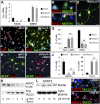
KHS101 specifically induces neuronal differentiation in rat NPCs. (A) Chemical structure of KHS101. (B) Real time RT-PCR analysis of NPCs treated with KHS101 for 24 h showing a dose-dependent induction of NeuroD mRNA expression normalized to the DMSO control. (C and D) TuJ1 staining (green) of DMSO- (0.1%, C) and KHS101-treated (5 μM, D) NPCs after a 4-d differentiation period (see also Figs. S1 and S2). (E and F) GFAP (green) and TuJ1 staining (red) of NPCs cultured under astrocyte-inducing conditions (50 ng/mL BMP4) in presence of DMSO (0.1%, J) or KHS101 (5 μM, K). (G) GFAP+ and TuJ1+ cell percentages show that KHS101 significantly suppresses BMP4-induced astrogenesis in NPCs, while significantly increasing neurogenesis. (H) Ingenuity pathway analysis reveals KHS101-induced up-regulation (red) of negative cell cycle regulators (Cdkn1 and Gadd45) and down-regulation of positive cell cycle regulators at 24 h of treatment (e.g., Ccnb1 and Ccne1; see also Fig. S3). (I) Real time RT-PCR analysis of NPCs treated with KHS101 for 24 h showing a dose-dependent induction of Cdkn1 mRNA expression, whereas inactive derivatives (KHS91, KHS92, and KHS103) fail to up-regulate Cdkn1 mRNA levels normalized to DMSO. (J and K) Ki67 (green) and P-HH3 staining (red) of DMSO- (0.1%, G) and KHS101-treated (5 μM, H) NPCs. (L) Ki67+ and P-HH3+ cell percentages indicate a significant reduction in proliferation and mitotic activity in KHS-treated NPCs over time. (Scale bars: 20 μm.) Error bars, SDs (three independent experiments; biological replicates in B and I); statistical significance (t test), *P < 0.05, **P < 0.01; nuclei were visualized with DAPI (blue).
Fig. 2.
Fig. 2.
KHS101 specifically interacts with TACC3 protein. (A) Structure of the benzophenone-containing alkyne-tagged KHS101 conjugate (KHS101-BP) used for target identification. (B) Representative two-dimensional SDS/PAGE and Western blotting of NPC cell lysates (2 mg/mL) detecting protein-KHS101-BP complexes after photocrosslinking (1 h) and biotin-tag labeling (25 μM biotin-azide, Left). Unlabeled KHS101 served as a competitor for specific KHS101-BP–protein binding (50-fold excess, Right). Independent experiments identified a protein spot that was reproducibly competed by unlabeled KHS101 (arrowheads). Mass spectrometry revealed the 80-kDa protein to be TACC3. (C) Western blot analysis of NPC lysate using a TACC3-specific antibody confirmed TACC3 identity after pulldown with KHS101-BP. (D) Recombinant rat TACC3 binds KHS101. Purified protein was incubated with biotinylated KHS101 (Table S1 and Schemes S3 and S4) in presence/absence of unlabeled compound, precipitated with streptavidin-coated agarose beads, and then detected by silver staining of SDS/PAGE gels.
Fig. 3.
Fig. 3.
Tacc3-specific shRNA recapitulates the neurogenic effect of KHS101 in rat NPCs. (A) Representative real time RT-PCR showing that Tacc3 RNA levels are markedly decreased in NPCs after electroporation with Tacc3-specific shRNA constructs (shTacc3-1, shTacc3-2). Concomitantly, Cdkn1 mRNA levels are elevated compared with the nontargeting control (shCO). (B and C) Nestin (green) and TACC3 (red) immunopositivity is observed in shCO-expressing NPCs (B, note centrosomal TACC3 localization) and strongly reduced upon TACC3 knockdown (C) (Fig. S4). (D) Tacc3-specific shRNA causes a TuJ1+ neuronal phenotype in NPCs. (E and F) Staining for Ki67 (red) and TuJ1 (green) in shCO- (E) and shTacc3-expressing NPCs (F) at 4 d after shRNA electroporation. (G) TuJ1+/Ki67− and Ki67+ cell percentages indicate significantly increased neurogenesis and significantly decreased proliferation upon Tacc3 RNAi in NPCs. (H and I) GFAP (green) and TuJ1 (red) staining in shCO- (H) and shTacc3-expressing (I) NPCs cultured with BMP4 (50 ng/mL) for 4 d. (J) GFAP+ and TuJ1+ cell percentages indicate that BMP4-induced astrogenesis is significantly reduced, whereas neurogenesis is significantly increased in NPCs upon Tacc3 RNAi. (K and L) Ectopic rat Tacc3 and Arnt2 cDNA overexpression in 293T cells and Western blot analysis. Increased expression of TACC3 is associated with decreased levels of nuclear ARNT2 (K). KHS101 treatment (0–15 μM) elevates nuclear but not cytoplasmic ARNT2 after 24 h of exposure (L). (M and N) Staining for ARNT2 reveals increased nuclear localization of ARNT2 in NPCs upon 5 μM KHS101 treatment for 12 h (Fig. S4). (Scale bars: 20 μm.) Error bars, SDs (three independent experiments; biological replicates in A); statistical significance (t test), *P < 0.05, **P < 0.01; nuclei were visualized with DAPI (blue).
Fig. 4.
Fig. 4.
KHS101 significantly increases neuronal differentiation in rats in vivo. (A) Pharmacokinetic profile of KHS101 in brain and plasma after single administration (3 mg/kg, i.v.) to Sprague–Dawley rats. (B and C) Immunohistochemistry of BrdU (red) and NeuN (green). White arrowheads mark BrdU-positive nuclei in the subgranular layer (SGL) of vehicle- and KHS101-treated animals. The yellow arrowhead marks a BrdU/NeuN double-positive nucleus, indicative of neuronal differentiation in the granule cell layer (GCL). (Scale bar: 20 μm.) (D and E) Ki67 (black arrowhead) and cleaved caspase 3 (brown arrowhead) double-stained DG sections (including the Hilus area: H) of vehicle- and KHS101-treated animals (E). (Scale bar: 50 μm.) (FH) Percentage of BrdU+/NeuN+ cells indicates significantly increased neurogenesis (F), whereas the number of Ki67-positive cells (G) significantly decreases in the DG upon KHS101 dosing; the number of cleaved caspase 3+ cells within the DG was not altered. (H) Quantification of BrdU+ cells localized in SGL, GL, and H of vehicle- and KHS101-treated animals (≥10 sections representative for the DG were counted for each animal). Note a significant reduction of BrdU+ cells in the SGL and a slight increase of BrdU+ cells in the GCL upon KHS101 administration. Error bars, SDs (six animals per group); statistical significance (t test), **P < 0.01.

References

    1. Zhao C, Deng W, Gage FH. Mechanisms and functional implications of adult neurogenesis. Cell. 2008;132:645–660. - PubMed
    1. Chan JP, Cordeira J, Calderon GA, Iyer LK, Rios M. Depletion of central BDNF in mice impedes terminal differentiation of new granule neurons in the adult hippocampus. Mol Cell Neurosci. 2008;39:372–383. - PMC - PubMed
    1. Hsieh J, Nakashima K, Kuwabara T, Mejia E, Gage FH. Histone deacetylase inhibition-mediated neuronal differentiation of multipotent adult neural progenitor cells. Proc Natl Acad Sci USA. 2004;101:16659–16664. - PMC - PubMed
    1. Kuwabara T, et al. Wnt-mediated activation of NeuroD1 and retro-elements during adult neurogenesis. Nat Neurosci. 2009;12:1097–1105. - PMC - PubMed
    1. Malberg JE, Duman RS. Cell proliferation in adult hippocampus is decreased by inescapable stress: Reversal by fluoxetine treatment. Neuropsychopharmacology. 2003;28:1562–1571. - PubMed

Publication types

MeSH terms

Associated data