Hippocampal neurons respond uniquely to topographies of various sizes and shapes
- PMID: 20823503
- PMCID: PMC4648552
- DOI: 10.1088/1758-5082/2/3/035005
Hippocampal neurons respond uniquely to topographies of various sizes and shapes
Abstract
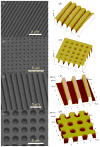
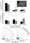
A number of studies have investigated the behavior of neurons on microfabricated topography for the purpose of developing interfaces for use in neural engineering applications. However, there have been few studies simultaneously exploring the effects of topographies having various feature sizes and shapes on axon growth and polarization in the first 24 h. Accordingly, here we investigated the effects of arrays of lines (ridge grooves) and holes of microscale (approximately 2 microm) and nanoscale (approximately 300 nm) dimensions, patterned in quartz (SiO2), on the (1) adhesion, (2) axon establishment (polarization), (3) axon length, (4) axon alignment and (5) cell morphology of rat embryonic hippocampal neurons, to study the response of the neurons to feature dimension and geometry. Neurons were analyzed using optical and scanning electron microscopy. The topographies were found to have a negligible effect on cell attachment but to cause a marked increase in axon polarization, occurring more frequently on sub-microscale features than on microscale features. Neurons were observed to form longer axons on lines than on holes and smooth surfaces; axons were either aligned parallel or perpendicular to the line features. An analysis of cell morphology indicated that the surface features impacted the morphologies of the soma, axon and growth cone. The results suggest that incorporating microscale and sub-microscale topographies on biomaterial surfaces may enhance the biomaterials' ability to modulate nerve development and regeneration.
Figures


Similar articles
-
Selective axonal growth of embryonic hippocampal neurons according to topographic features of various sizes and shapes.Int J Nanomedicine. 2010 Dec 22;6:45-57. doi: 10.2147/IJN.S12376. Int J Nanomedicine. 2010. PMID: 21289981 Free PMC article.
-
Immobilized nerve growth factor and microtopography have distinct effects on polarization versus axon elongation in hippocampal cells in culture.Biomaterials. 2007 Jan;28(2):271-84. doi: 10.1016/j.biomaterials.2006.07.043. Epub 2006 Aug 17. Biomaterials. 2007. PMID: 16919328
-
An electron microscopic analysis of hippocampal neurons developing in culture: early stages in the emergence of polarity.J Neurosci. 1993 Oct;13(10):4301-15. doi: 10.1523/JNEUROSCI.13-10-04301.1993. J Neurosci. 1993. PMID: 8410189 Free PMC article.
-
Biological responses to immobilized microscale and nanoscale surface topographies.Pharmacol Ther. 2018 Feb;182:33-55. doi: 10.1016/j.pharmthera.2017.07.009. Epub 2017 Jul 16. Pharmacol Ther. 2018. PMID: 28720431 Review.
-
Engineering microscale topographies to control the cell-substrate interface.Biomaterials. 2012 Jul;33(21):5230-46. doi: 10.1016/j.biomaterials.2012.03.079. Epub 2012 Apr 21. Biomaterials. 2012. PMID: 22521491 Free PMC article. Review.
Cited by
-
Recent advances in nanotherapeutic strategies for spinal cord injury repair.Adv Drug Deliv Rev. 2019 Aug;148:38-59. doi: 10.1016/j.addr.2018.12.011. Epub 2018 Dec 22. Adv Drug Deliv Rev. 2019. PMID: 30582938 Free PMC article. Review.
-
Engineered cell culture microenvironments for mechanobiology studies of brain neural cells.Front Bioeng Biotechnol. 2022 Dec 14;10:1096054. doi: 10.3389/fbioe.2022.1096054. eCollection 2022. Front Bioeng Biotechnol. 2022. PMID: 36588937 Free PMC article. Review.
-
Effect of Electrical and Electromechanical Stimulation on PC12 Cell Proliferation and Axon Outgrowth.Front Bioeng Biotechnol. 2021 Oct 21;9:757906. doi: 10.3389/fbioe.2021.757906. eCollection 2021. Front Bioeng Biotechnol. 2021. PMID: 34746110 Free PMC article.
-
Film-based Implants for Supporting Neuron-Electrode Integrated Interfaces for The Brain.Adv Funct Mater. 2014 Apr 2;24(13):1938-1948. doi: 10.1002/adfm.201303196. Adv Funct Mater. 2014. PMID: 25386113 Free PMC article.
-
Characterization of dorsal root ganglion neurons cultured on silicon micro-pillar substrates.Sci Rep. 2016 Dec 23;6:39560. doi: 10.1038/srep39560. Sci Rep. 2016. PMID: 28008963 Free PMC article.
References
-
- Li GN, Hoffman-Kim D. Tissue-engineered platforms of axon guidance. Tissue Eng B. 2008;14:33–51. - PubMed
-
- Liu CY, et al. Artificial niches for human adult neural stem cells: possibility for autologous transplantation therapy. J Hematother Stem Cell Res. 2003;12:689–99. - PubMed
-
- Norman J, Desai T. Methods for fabrication of nanoscale topography for tissue engineering scaffolds. Ann Biomed Eng. 2006;34:89–101. - PubMed
-
- Curtis A, Wilkinson C. Topographical control of cells. Biomaterials. 1997;18:1573–83. - PubMed
-
- Dalby MJ, Riehle MO, Yarwood SJ, Wilkinson CDW, Curtis ASG. Nucleus alignment and cell signaling in fibroblasts: response to a micro-grooved topography. Exp Cell Res. 2003;284:274–82. - PubMed
Publication types
MeSH terms
Grants and funding
LinkOut - more resources
Full Text Sources
