

A new crystal form of Lys48-linked diubiquitin
- PMID: 20823512
- PMCID: PMC2935213
- DOI: 10.1107/S1744309110027600
A new crystal form of Lys48-linked diubiquitin
Abstract
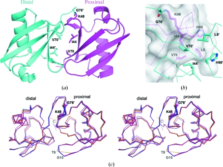
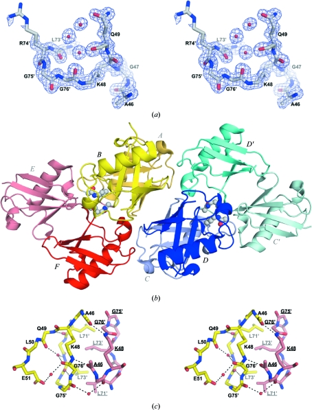
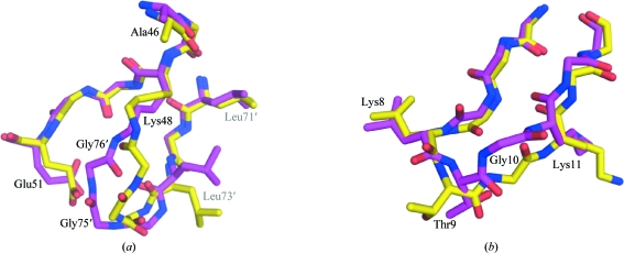
Lys48-linked polyubiquitin chains are recognized by the proteasome as a tag for the degradation of the attached substrates. Here, a new crystal form of Lys48-linked diubiquitin (Ub2) was obtained and the crystal structure was refined to 1.6 A resolution. The structure reveals an ordered isopeptide bond in a trans configuration. All three molecules in the asymmetric unit were in the same closed conformation, in which the hydrophobic patches of both the distal and the proximal moieties interact with each other. Despite the different crystallization conditions and different crystal packing, the new crystal structure of Ub2 is similar to the previously published structure of diubiquitin, but differences are observed in the conformation of the flexible isopeptide linkage.
Figures
Similar articles
-
Crystal structure of cyclic Lys48-linked tetraubiquitin.Biochem Biophys Res Commun. 2010 Sep 24;400(3):329-33. doi: 10.1016/j.bbrc.2010.08.057. Epub 2010 Aug 20. Biochem Biophys Res Commun. 2010. PMID: 20728431
-
Crystal structure and solution NMR studies of Lys48-linked tetraubiquitin at neutral pH.J Mol Biol. 2007 Mar 16;367(1):204-11. doi: 10.1016/j.jmb.2006.12.065. Epub 2006 Dec 29. J Mol Biol. 2007. PMID: 17240395
-
Structural and biochemical studies of the open state of Lys48-linked diubiquitin.Biochim Biophys Acta. 2012 Nov;1823(11):2046-56. doi: 10.1016/j.bbamcr.2012.04.003. Epub 2012 Apr 16. Biochim Biophys Acta. 2012. PMID: 22542781 Free PMC article.
-
Reading the ubiquitin postal code.Curr Opin Struct Biol. 2011 Dec;21(6):792-801. doi: 10.1016/j.sbi.2011.09.009. Epub 2011 Oct 27. Curr Opin Struct Biol. 2011. PMID: 22036065 Review.
-
[Structure of ubiquitin-recognition motifs and higher structures of polyubiquitin chains].Tanpakushitsu Kakusan Koso. 2005 Aug;50(10 Suppl):1311-21. Tanpakushitsu Kakusan Koso. 2005. PMID: 16104600 Review. Japanese. No abstract available.
Cited by
-
Ubiquitin and its binding domains.Front Biosci (Landmark Ed). 2012 Jun 1;17(6):2140-57. doi: 10.2741/4042. Front Biosci (Landmark Ed). 2012. PMID: 22652769 Free PMC article. Review.
-
Assessing the potential of atomistic molecular dynamics simulations to probe reversible protein-protein recognition and binding.Sci Rep. 2015 May 29;5:10549. doi: 10.1038/srep10549. Sci Rep. 2015. PMID: 26023027 Free PMC article.
-
The pros and cons of ubiquitination on the formation of protein condensates.Acta Biochim Biophys Sin (Shanghai). 2023 Jun 9;55(7):1084-1098. doi: 10.3724/abbs.2023096. Acta Biochim Biophys Sin (Shanghai). 2023. PMID: 37294105 Free PMC article.
-
Conformational dynamics of wild-type Lys-48-linked diubiquitin in solution.J Biol Chem. 2011 Oct 28;286(43):37496-502. doi: 10.1074/jbc.M111.256354. Epub 2011 Sep 7. J Biol Chem. 2011. PMID: 21900242 Free PMC article.
-
Comparison of native and non-native ubiquitin oligomers reveals analogous structures and reactivities.Protein Sci. 2016 Feb;25(2):456-71. doi: 10.1002/pro.2834. Epub 2016 Jan 12. Protein Sci. 2016. PMID: 26506216 Free PMC article.
References
-
- Bertolaet, B. L., Clarke, D. J., Wolff, M., Watson, M. H., Henze, M., Divita, G. & Reed, S. I. (2001). Nature Struct. Biol.8, 417–422. - PubMed
-
- Chen, Z. & Pickart, C. M. (1990). J. Biol. Chem.265, 21835–21842. - PubMed
-
- Cook, W. J., Jeffrey, L. C., Carson, M., Chen, Z. & Pickart, C. M. (1992). J. Biol. Chem.267, 16467–16471. - PubMed
-
- Cook, W. J., Jeffrey, L. C., Kasperek, E. & Pickart, C. M. (1994). J. Biol. Chem.236, 601–609. - PubMed
-
- Deveraux, Q., Ustrell, V., Pickart, C. & Rechsteiner, M. (1994). J. Biol. Chem.269, 7059–7061. - PubMed
Publication types
MeSH terms
Substances
Associated data
- Actions
Grants and funding
LinkOut - more resources
Full Text Sources
