

Carcinoma in situ testis displays permissive chromatin modifications similar to immature foetal germ cells
- PMID: 20823885
- PMCID: PMC2967056
- DOI: 10.1038/sj.bjc.6605880
Carcinoma in situ testis displays permissive chromatin modifications similar to immature foetal germ cells
Abstract
Background: The majority of testicular germ cell cancers develop through a pre-invasive carcinoma in situ (CIS) stage. The CIS cell is a neoplastic counterpart of foetal germ cells. During their development, foetal germ cells undergo extensive and essential epigenetic modifications, but little is known about epigenetic patterns in CIS cells.
Methods: Immunohistochemistry was used to investigate epigenetic patterns in CIS, germ cell tumours, normal adult and foetal testicular tissue.
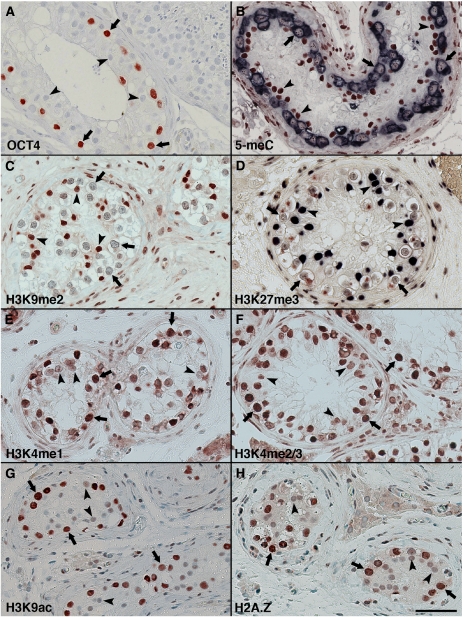
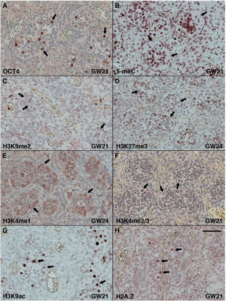
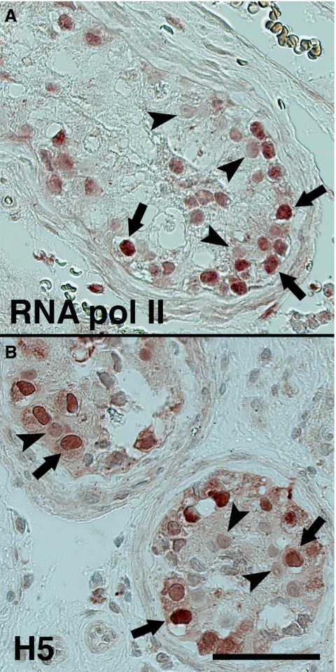
Results: CIS cells show low levels of DNA methylation and repressive histone modifications H3K9me2 and H3K27me3, but high levels of H3K9 acetylation, H3K4 methylation and H2A.Z, which all are associated with an activated and accessible chromatin structure. Collectively this renders a permissive chromatin structure and in accordance high levels of RNA polymerase II activity and proliferation (Ki-67 and mitotic index) is observed in CIS cells. Epigenetic patterns similar to that of CIS cells were observed in human gonocytes present within sex cords in foetal testes but correspond to migrating primordial germ cell in mice. Development of overt tumours involves epigenetic repression of the chromatin.
Conclusion: CIS cells have a permissive and foetal-like chromatin structure, which is associated with a high transcriptional and proliferative activity, likely empowering neoplastic transformation. Developmental epigenetic cues in foetal germ cells are substantially different between humans and mice.
Figures
References
-
- Agger K, Cloos P, Christensen J, Pasini D, Rose S, Rappsilber J, Issaeva I, Canaani E, Salcini A, Helin K (2007) UTX and JMJD3 are histone H3K27 demethylases involved in HOX gene regulation and development. Nature 449(7163): 731–734 - PubMed
-
- Albrechtsen R, Nielsen M, Skakkebaek N, Wewer U (1982) Carcinoma in situ of the testis. Some ultrastructural characteristics of germ cells. Acta Pathol Microbiol Immunol Scand A 90(4): 301–303 - PubMed
-
- Almstrup K, Hoei-Hansen C, Wirkner U, Blake J, Schwager C, Ansorge W, Nielsen J, Skakkebaek N, Rajpert-De Meyts E, Leffers H (2004) Embryonic stem cell-like features of testicular carcinoma in situ revealed by genome-wide gene expression profiling. Cancer Res 64(14): 4736–4743 - PubMed
-
- Barski A, Cuddapah S, Cui K, Roh T, Schones D, Wang Z, Wei G, Chepelev I, Zhao K (2007) High-resolution profiling of histone methylations in the human genome. Cell 129(4): 823–837 - PubMed
Publication types
MeSH terms
Substances
LinkOut - more resources
Full Text Sources
Medical
