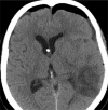
Incidence, radiographical features, and proposed mechanism for pneumocephalus from intravenous injection of air
- PMID: 20823969
- PMCID: PMC2908654
Incidence, radiographical features, and proposed mechanism for pneumocephalus from intravenous injection of air
Abstract
Background: Pneumocephalus typically implies a traumatic breach in the meningeal layer or an intracranial gas-producing infection. Unexplained pneumocephalus on a head computed tomography (CT) in an emergency setting often compels emergency physicians to undertake aggressive evaluation and consultation.
Methods: In this paper, we report three cases of pneumocephalus that appear to result from retrograde injection of air through an intravenous (IV) catheter. We also performed a retrospective study to determine the incidence of presumed IV-induced pneumocephalus and etiologies of pneumocephalus in our emergency department (ED) population.
Results: The incidence of idiopathic and presumed IV-induced pneumocephalus was 0.034% among all head CTs ordered in the ED and 4.88% among cases of pneumocephalus seen in the ED. These cases are characterized clinically by the absence of signs and symptoms of pathologic pneumocephalus and radiographically by the distribution of air densities along the cranial venous system on head CTs.
Conclusion: Idiopathic and presumed IV-induced pneumocephalus could be considered in the workup of ED patients with unexplained intracranial air on head CT if there are no findings of pathological causes for the pneumocephalus on history and physical examination and if the head CTs show a characteristic distribution of air limited to the cranial venous system. Knowledge of this clinical entity in the evaluation of ED patients with unexplained pneumocephalus can lead to more efficient emergency care and less patient anxiety.
Figures


References
-
- Counselman FL, Derkum S. An unusual cause of altered mental status. J Emerg Med. 1995;13(6):757–63. - PubMed
-
- Jelsma F, Moore DF. Cranial aerocele. Am J Surg. 1954;87:437–51. - PubMed
-
- Dandy WE. Pneumocephalus (Intracranial Pneumatocele or Aerocele) Archives of Surgery. 1926;12(5):949–82.
-
- Chan YP, Yau CY, Lewis RR, et al. Acute confusion secondary to pneumocephalus in an elderly patient. Age Ageing. 2000;29(4):365–7. - PubMed
-
- Markham JW. The clinical features of pneumocephalus based upon a survey of 284 cases with report of 11 additional cases. Acta Neurochir (Wien) 1967;16(1):1–78. - PubMed

