. 2010 Aug 24;1(3):e00178-10.
doi: 10.1128/mBio.00178-10.

Emergence and global dissemination of host-specific Streptococcus agalactiae clones

Affiliations

Emergence and global dissemination of host-specific Streptococcus agalactiae clones

Uffe B Skov Sørensen et al. mBio. .

Abstract

To examine the global diversity of Streptococcus agalactiae (group B streptococci [GBS]) and to elucidate the evolutionary processes that determine its population genetics structure and the reported changes in host tropism and infection epidemiology, we examined a collection of 238 bovine and human isolates from nine countries on five continents. Phylogenetic analysis based on the sequences of 15 housekeeping genes combined with patterns of virulence-associated traits identified a genetically heterogeneous core population from which virulent lineages occasionally emerge as a result of recombination affecting major segments of the genome. Such lineages, like clonal complex 17 (CC17) and two distinct clusters of CC23, are exclusively adapted to either humans or cattle and successfully spread globally. The recent emergence and expansion of the human-associated and highly virulent sequence type 17 (ST17) could conceivably account, in part, for the increased prevalence of neonatal GBS infections after 1960. The composite structure of the S. agalactiae genome invalidates phylogenetic inferences exclusively based on multilocus sequence typing (MLST) data and thereby the previously reported conclusion that the human-associated CC17 emerged from the bovine-associated CC67.


Figures
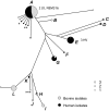
FIG 1
FIG 1
Preliminary clustering of bovine and human GBS isolates. The tree was constructed using the minimum evolution criterion. The analysis was based on concatemers generated from sequences of internal fragments of two genes, infB and sodA. A total of 238 GBS isolates were included in the analysis; of these, 228 were assigned to groups (groups A to L) of two or more isolates, and 10 were singletons (indicated by the smallest circles). The size of the circle reflects the number of isolates in the group. The sizes of groups consisting of one isolate (singleton), two isolates, and three isolates are shown in the symbol key. HV and LV indicate the locations of five serotype III isolates representing high-virulence and low-virulence clones (three HV clones and two LV clones), respectively, described by Musser et al. (11). Summaries of characteristics for isolates assigned to the 12 groups are listed in Table S2 in the supplemental material. The 55 representative isolates were selected within the groups (52 isolates) and among “outsiders” (three isolates marked by asterisks).
FIG 2
FIG 2
Schematic map of the S. agalactiae genome showing the positions of pilus islands (PI-1 and PI-2) and of genes examined in the present study. Genes used in conventional MLST analysis are indicated in green. The eight additional genes are indicated in red. Other genes, which were not included in the clustering analyses, are shown in blue. Locus tags and positions are given for the genes of strain 2603V/R (25), except for the genes missing in this GBS strain: those encoding β-antigen (bac) and 6-phospho-beta-galactosidase (lacG). The locus tags and positions for these two genes are given for strains A909 and NEM316, respectively. The complete genome sequences of the GBS strains NEM316, 2603V/R, and A909 have been determined (24–26), and all three show conserved gene order (synteny).
FIG 3
FIG 3
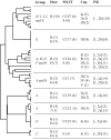
Extended characterization of 55 representative S. agalactiae isolates. The dendrogram shown to the left was constructed by a clustering analysis using the minimum evolution criterion based on concatemers generated from sequenced internal fragments of 15 genes. The table to the right of the dendrogram summarizes the main results for each cluster. For details, see Fig. S1 in the supplemental material. The table shows the provisional group assignment (Fig. 1; see Fig. S1 and Table S2 in the supplemental material), host, MLST result, cps type (Cap), and pilus island distribution. The host is indicated as follows: B, bovine; H, human. The clonal complex (e.g., CC67) and the presence of singletons (S) inferred by conventional MLST are shown in the MLST column. The pilus island distribution (Pili column) is indicated within brackets as follows: 1, presence of PI-1; [minus], absence of PI-1; 2a, presence of PI-2a; 2b, presence of PI-2b. The number of isolates is shown in parentheses in the host, MLST, Cap, and Pili columns.
FIG 4
FIG 4
Discordance between topologies of two trees obtained for the same set of GBS isolates based on MLSA schemes, including genes located in different genome sectors. The neighbor-joining tree obtained for the 55 representative GBS isolates using an MLSA scheme based on concatemers of seven genes located in sequential order in the last half of the genome (A) was compared with the tree obtained from the same strains using an MLSA scheme based on concatemers of eight genes located in sequential order in the first half of the genome (B). Relative genome positions of the genes included in the respective MLSA schemes are indicated in the genome diagrams. The strains were assigned to clonal complexes by MLST/eBURST analysis, and strain numbers were color coded accordingly. The average net distances between the clonal complexes are shown in the matrices. Distances were calculated from concatemers based on the two MLSA schemes. The numbers in parentheses are bootstrap values (1,000 replicates).

Similar articles

Cited by

References

    1. Elliott J. A., Facklam R. R., Richter C. B. 1990. Whole-cell protein patterns of nonhemolytic group B, type Ib, streptococci isolated from humans, mice, cattle, frogs, and fish. J. Clin. Microbiol. 28:628–630 - PMC - PubMed
    1. Keefe G. P. 1997. Streptococcus agalactiae mastitis: a review. Can. Vet. J. 38:429–437 - PMC - PubMed
    1. Merl K., Abdulmawjood A., Lämmler C., Zschock M. 2003. Determination of epidemiological relationships of Streptococcus agalactiae isolated from bovine mastitis. FEMS Microbiol. Lett. 226:87–92 - PubMed
    1. Anthony B. F., Okada D. M. 1977. The emergence of group B streptococci in infections of the newborn infant. Annu. Rev. Med. 28:355–369 - PubMed
    1. Trijbels-Smeulders M. A., Kollee L. A., Adriaanse A. H., Kimpen J. L., Gerards L. J. 2004. Neonatal group B streptococcal infection: incidence and strategies for prevention in Europe. Pediatr. Infect. Dis. J. 23:172–173 - PubMed

Publication types

LinkOut - more resources