. 2010 Sep 2;6(9):e1001087.
doi: 10.1371/journal.pgen.1001087.

Inhibition of GSK-3 ameliorates Abeta pathology in an adult-onset Drosophila model of Alzheimer's disease

Affiliations

Inhibition of GSK-3 ameliorates Abeta pathology in an adult-onset Drosophila model of Alzheimer's disease

Oyinkan Sofola et al. PLoS Genet. .

Erratum in

  • PLoS Genet. 2012 Jan;8(1). doi: 10.1371/annotation/baa8a2a9-130b-4959-b6fb-6f786fd02826

Abstract

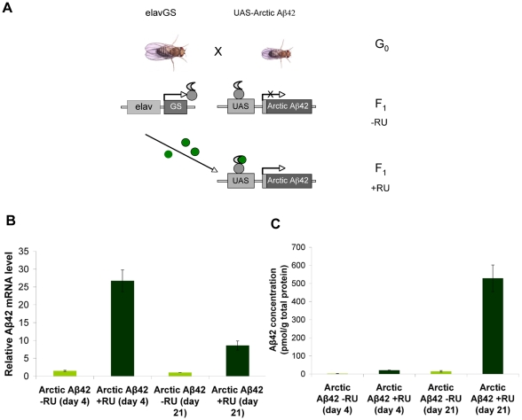
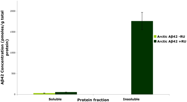
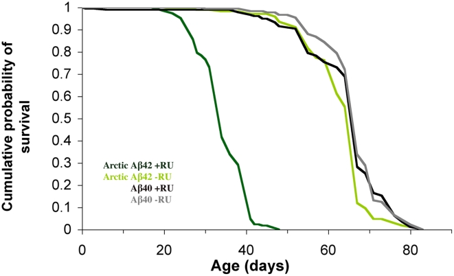
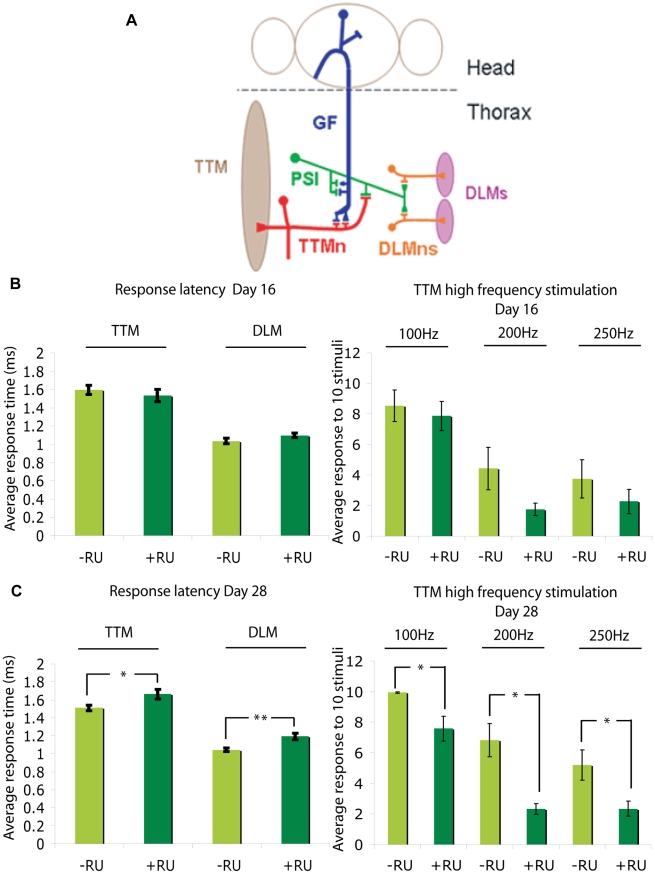
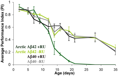
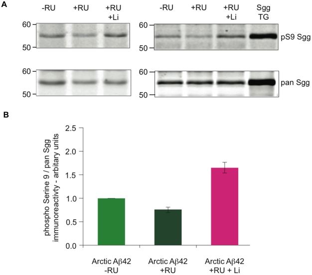
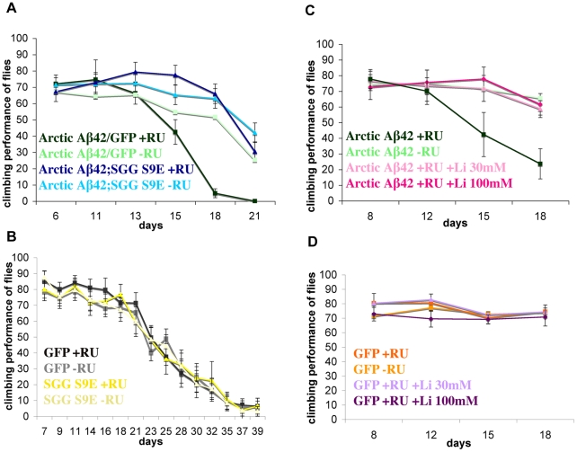
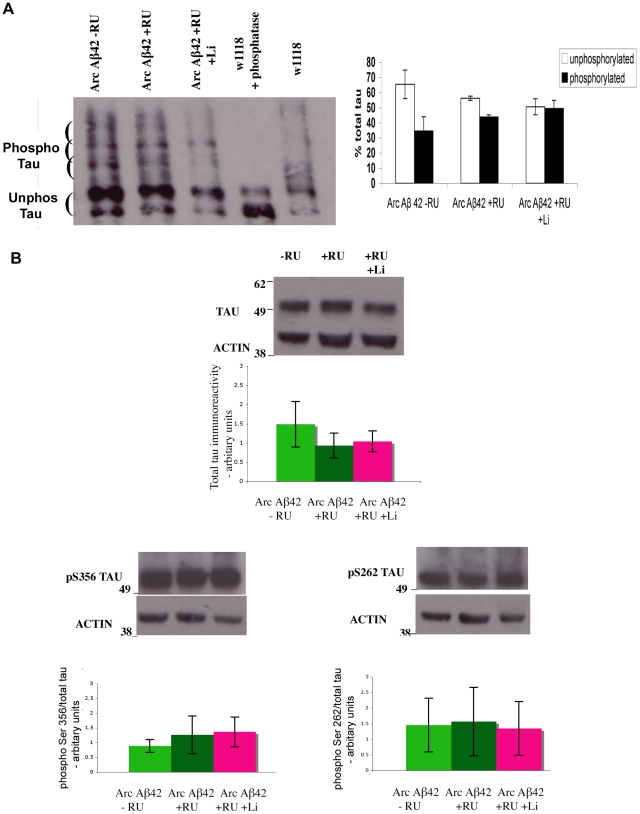
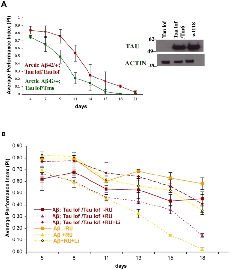
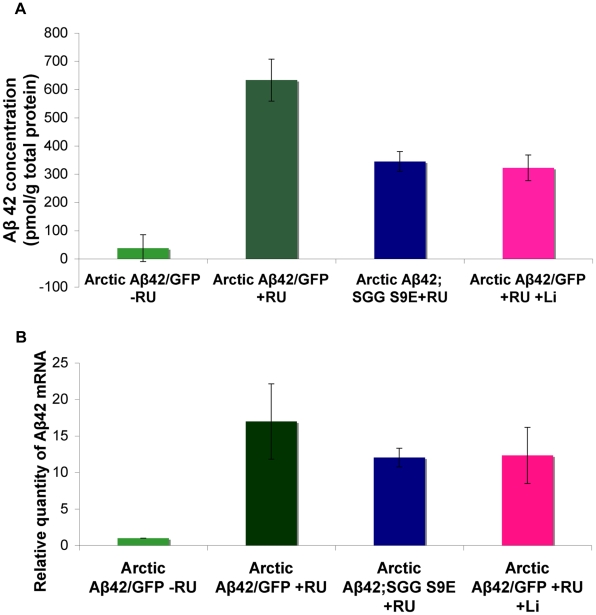
Abeta peptide accumulation is thought to be the primary event in the pathogenesis of Alzheimer's disease (AD), with downstream neurotoxic effects including the hyperphosphorylation of tau protein. Glycogen synthase kinase-3 (GSK-3) is increasingly implicated as playing a pivotal role in this amyloid cascade. We have developed an adult-onset Drosophila model of AD, using an inducible gene expression system to express Arctic mutant Abeta42 specifically in adult neurons, to avoid developmental effects. Abeta42 accumulated with age in these flies and they displayed increased mortality together with progressive neuronal dysfunction, but in the apparent absence of neuronal loss. This fly model can thus be used to examine the role of events during adulthood and early AD aetiology. Expression of Abeta42 in adult neurons increased GSK-3 activity, and inhibition of GSK-3 (either genetically or pharmacologically by lithium treatment) rescued Abeta42 toxicity. Abeta42 pathogenesis was also reduced by removal of endogenous fly tau; but, within the limits of detection of available methods, tau phosphorylation did not appear to be altered in flies expressing Abeta42. The GSK-3-mediated effects on Abeta42 toxicity appear to be at least in part mediated by tau-independent mechanisms, because the protective effect of lithium alone was greater than that of the removal of tau alone. Finally, Abeta42 levels were reduced upon GSK-3 inhibition, pointing to a direct role of GSK-3 in the regulation of Abeta42 peptide level, in the absence of APP processing. Our study points to the need both to identify the mechanisms by which GSK-3 modulates Abeta42 levels in the fly and to determine if similar mechanisms are present in mammals, and it supports the potential therapeutic use of GSK-3 inhibitors in AD.


Conflict of interest statement

The authors have declared that no competing interests exist.

Figures

Figure 1
Figure 1. Adult-onset induction of Arctic Aβ42 peptide in the Drosophila nervous system.
(A) A schematic representation of the GeneSwitch-UAS expression system (based on [30]). Driver lines expressing the transcriptional activator GeneSwitch under control of the nervous system-specific elav promoter (elavGS) are crossed to flies expressing an Aβ transgene fused to a GAL4-binding upstream activation sequence (UAS-Aβ). In the absence of the activator mifepristone (RU486; −RU), the GeneSwitch protein is expressed in neurons but remains transcriptionally silent, so that Aβ is not expressed. Following treatment with RU486 (+RU; green) the GeneSwitch protein is transcriptionally activated, binds to UAS and thus mediates expression of Aβ peptide specifically in the fly nervous system. Aβ42 RNA (B) and protein (C) levels were quantified at four days and 21 days post-RU486 treatment (see Materials and Methods). Data are presented as means ± SEM and were analysed by two-way ANOVA and Tukey's honestly significant difference (HSD) post-hoc comparisons. P<0.05 comparing Aβ RNA expression in RU486-treated UAS-ArcAβ42/+;elavGS/+ flies to their −RU486 controls at both time-points (Tukey's HSD). P<0.01 comparing Aβ42 protein levels in RU486-treated UAS-ArcAβ42/+;elavGS/+ flies to untreated controls at both time-points.
Figure 2
Figure 2. Arctic Aβ42 peptide in the adult Drosophila nervous system is mostly in an insoluble fibrillar state.
In the absence of the activator mifepristone (RU486; −RU), a negligible amount of soluble protein is observed at day 15. Following treatment with RU486 (+RU; dark green) the Aβ peptide expression is seen in both soluble and insoluble fractions with a significant proportion observed in the insoluble fraction. Data are presented as means ± SEM and were analysed by ANOVA, P<0.01 when protein levels of soluble and insoluble fractions of Aβ42 expressing flies were compared.
Figure 3
Figure 3. Expression of Arctic Aβ42 specifically in the adult nervous system shortens lifespan.
Lifespans were determined as described in materials & methods. Survival curves are depicted and data were compared using the log-rank test. P<0.01 comparing median lifespan of UAS-ArcAβ42/+;elavGS/+ +RU flies to their −RU controls. P<0.01 comparing UAS-ArcAβ42/+;elavGS/+ +RU flies to UAS-Aβ40/+;elavGS/+ +RU controls. Induction of Aβ40 in the adult nervous system did not alter lifespan in comparison to non-RU486-treated controls.
Figure 4
Figure 4. Arctic Aβ42 peptides induce progressive, adult-onset neuronal defects in Drosophila.
(A) A schematic illustration of the Drosophila giant fibre system (GFS; adapted from [74]). Giant fibres (GFs; blue) relay signals from the brain to the thoracic musculature via mixed electrochemical synapses with the motorneurons (TTMn, red) of the tergotrancheral muscle (TTM; left), and the peripherally synapsing interneuron (PSI; green), which subsequently forms chemical synapses with the motorneurons (DLMn; orange) of the dorsal longitudinal muscles (DLM; right). Note only one of the TTMn axons is shown exiting the nervous system and contacting the muscle on the left hand side and one set of the DLMns and corresponding neuromuscular junctions are depicted on the right hand side. GFS activity was measured in UAS-ArcAβ42;elavGS flies at (B) 16 days and (C) 28 days post-RU486 treatment (see Materials and Methods); parameters measured were the latencies from GF stimulation to muscle response (response latency DLM and TTM) and the stability of the response to high frequency stimulation at 100, 200 and 250 Hz (high frequency stimulation TTM). Data are presented as the mean response ± SEM and were analysed by student's t-test, at each time point, on log-derived data. *P<0.05, **P<0.01 comparing response latency or response to high frequency stimulation of UAS-ArcAβ42/+;elavGS/+ +RU flies to −RU controls at 28 days post-induction.
Figure 5
Figure 5. Expression of Arctic Aβ42 peptides in the adult fly nervous system causes locomotor dysfunction.
Climbing ability of UAS-ArcAβ42/+;elavGS/+ and UAS-Aβ40/+;elavGS/+ flies on + and − RU486 SY medium was assessed at the indicated time-points (see Materials and Methods). Data are presented as the average performance index (PI) ± SEM and were compared using two-way ANOVA and Tukey's honestly significant difference (HSD) post-hoc analyses (number of independent tests (n) = 3, number of flies per group (nt) = 39–45). *P<0.05 comparing PI of UAS-ArcAβ42/+;elavGS/+ +RU486 flies to that of untreated and Aβ40 over-expressing controls at the indicated time points (Tukey's HSD).
Figure 6
Figure 6. Flies expressing Aβ42 in adult neurons show a decrease in Shaggy inhibitory Ser9 phosphorylation.
(A) Western blot analyses for pan-Sgg and phospho Ser9 Sgg revealed a decrease in Ser9 phosphorylation in flies expressing Aβ42 by RU induction for 15 days (UAS-ArcAβ42/+;elavGS/+ or UAS-ArcAβ42/GFP;elavGS/+) in comparison to their −RU controls, while an increase in Ser9 phosphorylation was observed when the Aβ42 expressing flies were fed lithium (+RU +Li). Flies over expressing Sgg were used as positive control. (B) Quantification of the western blot analysis in (A), n = 3, is depicted in the bar chart, with significant differences seen between ArcAβ42/+;elavGS/+ +RU flies to −RU controls (P<0.01), and between ArcAβ42/+;elavGS/+ +RU +Li flies to +RU controls (P<0.001).
Figure 7
Figure 7. Expression of the dominant negative mutant Shaggy S9E in the adult nervous system extends the median and maximum lifespan of Arctic Aβ42 flies.
(A). Survival curves of flies co-expressing Arctic Aβ42 and SggS9E are depicted and data were compared using the log-rank test. P<0.001 comparing UAS-ArcAβ42/+;elavGS/UAS-SggS9E +RU flies to UAS-ArcAβ42/UAS-gfp;elavGS/+ +RU controls. No significant difference was seen in the −RU controls. (B). Expression of Shaggy S9E alone did not affect control lifespan. No significant difference was observed comparing elavGS/UAS-SggS9E +RU to either elavGS/UAS-SggS9E −RU, or UAS-gfp/elavGS/+ +RU control lifespans.
Figure 8
Figure 8. Inhibition of Shaggy, either by expression of SggS9E in the adult nervous system or treatment with lithium, suppresses the locomotor dysfunction phenotype of Arctic Aβ42 flies.
(A) Climbing ability of UAS-ArcAβ42/UAS-gfp;elavGS/+ and UAS-ArcAβ42/+;elavGS/UAS-SggS9E flies on +RU486 SY medium was assessed at the indicated time-points (see Materials and Methods). Data are presented as the percentage climbing performance of flies ± SD. P<0.001 when UAS-ArcAβ42/UAS-gfp;elavGS/+ and UAS-ArcAβ42/+;elavGS/UAS-SggS9E flies are compared at day 21 (one-way ANOVA, number of independent tests (n) = 3). Graph shows one representative data of repeated experiments. (B) Expression of Shaggy S9E alone does not reduce climbing ability of control flies. elavGS/UAS-SggS9E +RU flies display a similar locomotor function compared to both elavGS/UAS-SggS9E −RU and UAS-gfp/+;elavGS/+ +RU control flies. (C) Climbing ability of UAS-ArcAβ42/+;elavGS/+ and UAS-ArcAβ42/UAS-gfp;elavGS/+ on +RU486 SY medium was assessed in the presence and absence of lithium chloride (30mM and 100mM). P<0.001 when UAS-ArcAβ42 flies were fed lithium and compared to flies not fed lithium at day 18 (one-way ANOVA, n = 3). Graph shows one representative data of repeated experiments. (D) Lithium had no effect on negative geotaxis of control flies. UAS-gfp/+;elavGS/+ +RU and UAS-gfp/+;elavGS/+ +RU in the presence of lithium had similar locomotor function. The crosses were performed at 27°C.
Figure 9
Figure 9. Flies expressing Aβ42 in adult neurons show no changes in total tau phosphorylation and at Ser 262 and Ser 356 specific epitopes.
(A) Western blot analyses for total tau phosphorylation using phos-tag gels and (B) phospho Ser262 and Ser356. Tau showed no changes in phosphorylation in flies expressing Aβ42 (UAS-ArcAβ42/GFP;elavGS/+) in comparison to their −RU controls, and when the Aβ42 expressing flies were fed lithium (+RU +Li). Flies were collected 17 days post RU induction, and maintained at 27°C. Quantification of the western blot analysis in (A), phospho-tau or non-phospho-tau normalised to total tau per sample (n = 4), and in (B) n = 4 for total tau and n = 3 for the Ser sites depicted in the bar chart, showed no significant differences between ArcAβ42/+;elavGS/+ +RU flies to −RU controls or to ArcAβ42/+;elavGS/+ +RU +Li flies (one-way ANOVA).
Figure 10
Figure 10. Lithium treatment alone has a greater protective effect against Aβ42 toxicity than loss of tau function alone.
(A) Loss of tau partially suppressed the locomotor dysfunction phenotype of Arctic Aβ42 flies. Expression of Arctic Aβ42 peptide in a tau heterozygous background in the adult fly nervous system caused locomotor dysfunction. This phenotype was suppressed when Arctic Aβ42 peptide was expressed in a homozygous tau mutant background. Climbing ability of UAS-ArcAβ42/+;elavGS Tau EP3203/TM6 and UAS-ArcAβ42/+;elavGS Tau EP3203/Tau Dfc flies on +RU486 SY medium was assessed at the indicated time-points (see Materials and Methods). Data are presented as the average performance index (PI) ± SEM (number of independent tests (n) = 3, number of flies per group (nt) = 45). P<0.0001 comparing the PI of UAS-ArcAβ42/+;elavGS Tau EP3203/Tau Dfc to UAS-ArcAβ42/+;elavGS Tau EP3203/TM6 flies (two-way ANOVA). The crosses were performed at 27°C. Western blotting analysis, using a non-phosphorylation dependent antibody to Drosophila tau, confirmed that endogenous tau protein levels were greatly reduced in UAS-ArcAβ42/+;elavGS tau EP3203/tau Dfc flies in comparison to tau heterozygous flies (UAS-ArcAβ42/+;elavGS tau EP3203/TM6) and control w1118 flies. (B) Arctic Aβ42 toxicity was suppressed when the peptide was expressed in a homozygous tau mutant background or when flies were treated with lithium. Lithium treatment alone had a greater protective effect against Aβ42 toxicity than did loss of tau function alone, and was not dependent on tau. Climbing ability of UAS-ArcAβ42/+;elavGS/+ and UAS-ArcAβ42/+;elavGS tau EP3203/tau Dfc flies on − or +RU486 SY medium, in the presence or absence of lithium, was assessed at the indicated time-points. Data are presented as the average PI ± SEM and were analysed by two-way ANOVA (number of independent tests (n) = 4, number of flies per group (nt) = 60). Comparing PI of UAS-ArcAβ42/UAS-gfp;elavGS/+ and UAS-ArcAβ42/+;elavGS tau EP3203/tau Dfc flies on −RU, significant differences between genotypes (P<0.0001) were observed. P = 0.0002 comparing PI of UAS-ArcAβ42/UAS-gfp;elavGS/+ and UAS-ArcAβ42/+;elavGS tau EP3203/tau Dfc flies on +RU food. No significant differences were observed comparing UAS-ArcAβ42/+;elavGS/+ and UAS-ArcAβ42/+;elavGS tau EP3203/tau Dfc flies on +RU +lithium (P = 0.692).
Figure 11
Figure 11. Inhibiting Shaggy activity reduces amyloid levels of Arctic Aβ42 flies.
(A) Protein levels of UAS-ArcAβ42/UAS-gfp;elavGS/+ and UAS-ArcAβ42/+;elavGS/UAS-SggS9E flies on +RU486 SY medium, and UAS-ArcAβ42/UAS-gfp;elavGS/+ flies on +RU +Lithium (Li), were measured by ELISA at 15 days post-induction (see Materials and Methods). Data were compared using one-way ANOVA, number of independent tests (n) = 3. P<0.01, and P<0.0001 when comparing UAS-ArcAβ42/UAS-gfp;elavGS/+ to UAS-ArcAβ42/+;elavGS/UAS-SggS9E flies on +RU486 SY medium, and UAS-ArcAβ42/UAS-gfp;elavGS/+ +RU to UAS-ArcAβ42/UAS-gfp;elavGS/+ +RU +Li respectively. (B) RNA levels of UAS-ArcAβ42/UAS-gfp;elavGS/+, UAS-ArcAβ42/+;elavGS/UAS-SggS9E +RU486 SY medium and UAS-ArcAβ42/UAS-gfp;elavGS/+ +RU + lithium flies were measured at day 5. No significant difference was seen in the levels of RNA expression, number of independent tests (n) = 3.

References

    1. Spires TL, Hyman BT. Transgenic models of Alzheimer's disease: learning from animals. NeuroRx. 2005;2:423–437. - PMC - PubMed
    1. Selkoe DJ, Schenk D. Alzheimer's disease: molecular understanding predicts amyloid-based therapeutics. Annu Rev Pharmacol Toxicol. 2003;43:545–584. - PubMed
    1. Hutton M, Lendon CL, Rizzu P, Baker M, Froelich S, et al. Association of missense and 5′-splice-site mutations in tau with the inherited dementia FTDP-17. Nature. 1998;393:702–705. - PubMed
    1. Rizzu P, Van Swieten JC, Joosse M, Hasegawa M, Stevens M, et al. High prevalence of mutations in the microtubule-associated protein tau in a population study of frontotemporal dementia in the Netherlands. Am J Hum Genet. 1999;64:414–421. - PMC - PubMed
    1. Lewis J, Dickson DW, Lin WL, Chisholm L, Corral A, et al. Enhanced neurofibrillary degeneration in transgenic mice expressing mutant tau and APP. Science. 2001;293:1487–1491. - PubMed

Publication types

MeSH terms

LinkOut - more resources