. 2010 Sep 1;5(9):e12500.
doi: 10.1371/journal.pone.0012500.

Gene knock-outs of inositol 1,4,5-trisphosphate receptors types 1 and 2 result in perturbation of cardiogenesis

Affiliations

Gene knock-outs of inositol 1,4,5-trisphosphate receptors types 1 and 2 result in perturbation of cardiogenesis

Keiko Uchida et al. PLoS One. .

Abstract

Background: Inositol 1,4,5-trisphosphate receptors (IP3R1, 2, and 3) are intracellular Ca2+ release channels that regulate various vital processes. Although the ryanodine receptor type 2, another type of intracellular Ca2+ release channel, has been shown to play a role in embryonic cardiomyocytes, the functions of the IP3Rs in cardiogenesis remain unclear.

Methodology/principal findings: We found that IP3R1(-/-)-IP3R2(-/-) double-mutant mice died in utero with developmental defects of the ventricular myocardium and atrioventricular (AV) canal of the heart by embryonic day (E) 11.5, even though no cardiac defect was detectable in IP3R1(-/-) or IP3R2(-/-) single-mutant mice at this developmental stage. The double-mutant phenotype resembled that of mice deficient for calcineurin/NFATc signaling, and NFATc was inactive in embryonic hearts from the double knockout-mutant mice. The double mutation of IP3R1/R2 and pharmacologic inhibition of IP3Rs mimicked the phenotype of the AV valve defect that result from the inhibition of calcineurin, and it could be rescued by constitutively active calcineurin.

Conclusions/significance: Our results suggest an essential role for IP3Rs in cardiogenesis in part through the regulation of calcineurin-NFAT signaling.


Conflict of interest statement

Competing Interests: The authors have declared that no competing interests exist.

Figures

Figure 1
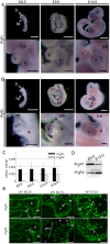
Figure 1. Both IP3R1 and IP3R2 are expressed in the embryonic heart.
(A, B) Whole-mount in situ hybridization using IP3R1 (A) and IP3R2 (B) antisense riboprobes at E8.5, E9.5, and E10.5. Whole-mount views (upper panels) and close-up views of the hearts (lower panels) are shown. Scale bars: 1 mm (whole-mount views) and 0.2 mm (heart close-up views). (C) Quantitative RT-PCR of IP3R1 (black bars) and IP3R2 (gray bars) using total RNA samples extracted from embryonic hearts at E8.5 to E16.0. Error bars indicate standard deviations. (D) Western blot of embryonic hearts with anti-IP3R1 (18A10) and anti-IP3R2 (KM1083) antibodies. Lysates (10 µg) of hearts at E9.5 and E12.5 were analyzed. The protein contents of the heart extracts were determined by the Bradford method. (E) The transverse sections of the wildtype embryos at E9.25, E9.75 and E10.5 were immunostained with the anti-IP3R1 (upper panels) and anti-IP3R2 (lower panels) antibodies. Scale bars: 0.1 mm. a, atrium; avc, atrioventricular canal; e, endocardium; h, head; ht, heart; m, myocardium; pa, pharyngeal arch; v, ventricle.
Figure 2
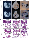
Figure 2. Cardiac defects in IP3R1−/−-IP3R2−/− mice.
(A) The morphologies of the embryos and developing hearts of IP3R1+/−-IP3R2−/− (upper panels) and IP3R1−/−-IP3R2−/− (lower panels) mice at E9.25 and at E9.75. Scale bars, 0.5 mm (whole-mount views) and 0.2 mm (heart close-up views). (B) Hematoxylin and eosin-stained transverse sections of the anterior, middle and posterior segments of the hearts of IP3R1+/−-IP3R2−/− (upper panels) and IP3R1−/−-IP3R2−/− (lower panels) mice at E9.75. The insets in the middle panels are higher-magnification images of the boxed areas. Scale bars, 0.2 mm. a, atrium; avc, atrioventricular canal; fg, foregut; h, head; ht, heart; lv, left ventricle; oft, outflow tract; pa, pharyngeal arch; ph, pharynx; rv, right ventricle.
Figure 3
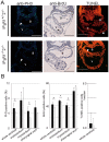
Figure 3. IP3R1 and IP3R2 redundantly regulate cardiomyocyte proliferation in the developing ventricles.
(A) Transverse sections of E9.5 IP3R1+/−-IP3R2−/− (upper panels) and IP3R1−/−-IP3R2−/− (lower panels) embryos that were immunostained with the anti-phospho-histone H3 (PH3) antibody, immunostained with the anti-BrdU antibody after injection of BrdU, and subjected to the TUNEL assay. The green signals (white arrowheads) in the left panels, brown signals in the middle panels (arrows) and yellow signals (white arrowheads) in the right panels indicate the nuclei of the PH3-, BrdU-, and TUNEL-positive cells, respectively. Scale bars, 0.2 mm. a, atrium; v, ventricle. (B) The left and middle graphs show the percentages of proliferating cells in the whole ventricles, the endocardium and myocardium of the ventricles, and the pharyngeal arches of IP3R1−/−-IP3R2−/− mutants (gray bars), compared with those of IP3R1+/−-IP3R2−/− controls (black bars) (*P<0.05, n = 3). The right graph shows the number of apoptotic cells (P = 0.45, n = 7). Error bars indicate standard deviations.
Figure 4
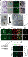
Figure 4. IP3Rs are essential for EMT through calcineurin activity during endocardial cushion development in mice.
(A) Hematoxylin and eosin staining, alcian blue staining, staining with biotinylated hyaluronan binding protein and whole-mount in situ hybridization for a marker of the atrioventricular (AV) myocardium (Tbx2) of IP3R1+/−-IP3R2−/− (upper panels) and IP3R1−/−-IP3R2−/− (lower panels) embryos at E9.5. Mesenchymal cells are absent from the IP3R1−/−-IP3R2−/− AV cushion (arrowheads), whereas the staining levels for alcian blue (blue signals), hyaluronan (green signals) and Tbx2 (purple signals) are not altered. Scale bars, 0.1 mm. (B) In vitro EMT assay using the endocardial cushion from the AV canal. The left panels show that treatment of wild-type (WT) cushion explants with the IP3R inhibitor 2APB inhibits the outgrowth of spindle-shaped cells as compared with the control treatment (DMSO). The right panels show the IP3R1+/−-IP3R2−/− and the IP3R1−/−-IP3R2−/− AV cushion explants. The higher magnification views of the explants are shown besides. The spindle-shaped cells migrating into collagen gel are indicated with yellow arrowheads. The number of the spindle-shaped migrating cells is significantly lower in the culture that contains 2APB (left graph) and in the culture from the IP3R1−/−-IP3R2−/− AV cushion (right graph) (*P<0.05, n = 3). Error bars indicate standard deviations. (C) Transverse sections at the level of the AV canal stained with the anti-NFATc1 antibody and TOPRO3 show impairment of translocation of NFATc1 into nuclei at E9.5. The expression level of NFATc1 is decreased at E10.5 in IP3R1−/−-IP3R2−/− hearts. Scale bars, 0.05 mm. Arrowheads indicate the AV canal and higher-magnification images of the boxed area. (D) Western blot analysis with anti-NFATc4 antibody using heart lysates of the wildtype and the IP3R1−/−-IP3R2−/− embryos at E9.75. Blotting with anti-GAPDH antibody on each lane was used as a loading control. The inactive form of NFATc4 (p-NFATc4) remained and active form (NFATc4) was reduced in IP3R1−/−-IP3R2−/− hearts compared to IP3R1+/−-IP3R2−/− hearts. (E) The structures of the wildtype calcineurin (WT CN) and the constitutively active form of calcineurin (CA CN) cDNA are shown. Infection with CA CN cDNA resulted in significant increase in the number of the spindle-shaped and α smooth muscle actin (αSMA)-positive cells (yellow arrowheads) in the culture of IP3R1−/−-IP3R2−/− AV cushion explants (*P<0.05, n = 4). a, atrium; e, endocardium; lv, left ventricle; m, myocardium, rv, right ventricle; v, ventricle.
Figure 5
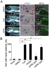
Figure 5. Inhibition of IP3Rs and calcineurin results in a common developmental defect in zebrafish hearts.
(A) The left panels show the gross abnormalities in the hearts of zebrafish treated with DMSO, CsA (calcineurin inhibitor), and 2APB (IP3R inhibitor). Atrioventricular regurgitation is induced by the addition of CsA and 2APB, resulting in heart failure with pericardial swelling (yellow arrowheads). The middle panels show the histologic abnormalities including a decrease in the number of cushion cells in the atrioventricular valves (arrowheads). The right panels show the expression of sarcomeric protein MF20 in zebrafish hearts that did not alter with CsA- or 2APB-treatment. (B) The significant increase in the number of zebrafish with pericardial swelling following the addition of CsA, FK506, or 2APB, is indicated in the graph (*P<0.05, n = 3). Error bars indicate standard deviations. a, atrium; ey, eye; ht, heart; v, ventricle; y, yolk.

References

    1. Berridge MJ, Bootman MD, Roderick HL. Calcium signalling: dynamics, homeostasis and remodelling. Nat Rev Mol Cell Biol. 2003;4:517–529. - PubMed
    1. Kockskamper J, Zima AV, Roderick HL, Pieske B, Blatter LA, et al. Emerging roles of inositol 1,4,5-trisphosphate signaling in cardiac myocytes. J Mol Cell Cardiol. 2008;45:128–147. - PMC - PubMed
    1. Furuichi T, Yoshikawa S, Miyawaki A, Wada K, Maeda N, et al. Primary structure and functional expression of the inositol 1,4,5-trisphosphate-binding protein P400. Nature. 1989;342:32–38. - PubMed
    1. Iwai M, Tateishi Y, Hattori M, Mizutani A, Nakamura T, et al. Molecular cloning of mouse type 2 and type 3 inositol 1,4,5-trisphosphate receptors and identification of a novel type 2 receptor splice variant. J Biol Chem. 2005;280:10305–10317. - PubMed
    1. Matsumoto M, Nakagawa T, Inoue T, Nagata E, Tanaka K, et al. Ataxia and epileptic seizures in mice lacking type 1 inositol 1,4,5-trisphosphate receptor. Nature. 1996;379:168–171. - PubMed

Publication types

Substances