The classic: The bonding of prostheses to bone by cement. 1964
- PMID: 20824402
- PMCID: PMC2974882
- DOI: 10.1007/s11999-010-1545-8
The classic: The bonding of prostheses to bone by cement. 1964
Abstract
This Classic Article is a reprint of the original work by Sir John Charnley, The Bonding of Prostheses to Bone by Cement. An accompanying biographical sketch of Sir John Charnley, MD is available at DOI 10.1007/s11999-010-1547-6. The Classic Article is reproduced and adapted with permission and copyright © of the British Editorial Society of Bone and Joint Surgery from Charnley J. The bonding of prostheses to bone by cement. J Bone Joint Surg [Br]. 1964;46-B:518–529.
Figures

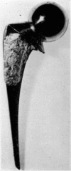
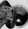
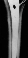
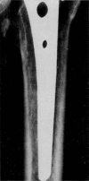
Similar articles
-
50 Years ago in CORR: Complications in replacement arthroplasty of the hip; experience with 68 additional cases Howard Mendelsohn, MD and Bernard N. Becker, MD CORR 1955;6:48-53.Clin Orthop Relat Res. 2010 Sep;468(9):2550-2. doi: 10.1007/s11999-010-1420-7. Clin Orthop Relat Res. 2010. PMID: 20544316 Free PMC article. No abstract available.
-
50 years ago in CORR: Biomechanics of hip prostheses. Duncan C. McKeever, MD CORR 1961;19:187-199.Clin Orthop Relat Res. 2012 Feb;470(2):626-7. doi: 10.1007/s11999-011-2195-1. Epub 2011 Nov 19. Clin Orthop Relat Res. 2012. PMID: 22101406 Free PMC article. No abstract available.
-
The Sivash constrained acetabular cup.Orthopedics. 2009 Mar;32(3):157. Orthopedics. 2009. PMID: 19309069 No abstract available.
-
Primary total hip arthroplasty.AORN J. 2003 Dec;78(6):947-53, 956-69; quiz 971-4. doi: 10.1016/s0001-2092(06)60586-3. AORN J. 2003. PMID: 14692668 Review.
-
Surface replacement arthroplasty of the hip.Bull NYU Hosp Jt Dis. 2009;67(1):75-82. Bull NYU Hosp Jt Dis. 2009. PMID: 19302061 Review.
Cited by
-
A promising material for bone repair: PMMA bone cement modified by dopamine-coated strontium-doped calcium polyphosphate particles.R Soc Open Sci. 2019 Oct 2;6(10):191028. doi: 10.1098/rsos.191028. eCollection 2019 Oct. R Soc Open Sci. 2019. PMID: 31824710 Free PMC article.
-
Dr. John H. Charnley: An Architect and Pioneer of the Modern Era of Hip Replacement Surgery.Cureus. 2024 Sep 6;16(9):e68832. doi: 10.7759/cureus.68832. eCollection 2024 Sep. Cureus. 2024. PMID: 39376811 Free PMC article. Review.
-
MiR-106b inhibition suppresses inflammatory bone destruction of wear debris-induced periprosthetic osteolysis in rats.J Cell Mol Med. 2020 Jul;24(13):7490-7503. doi: 10.1111/jcmm.15376. Epub 2020 Jun 2. J Cell Mol Med. 2020. PMID: 32485091 Free PMC article.
-
Linoleic acid addition prevents Staphylococcus aureus biofilm formation on PMMA bone cement.Biofilm. 2025 Aug 7;10:100311. doi: 10.1016/j.bioflm.2025.100311. eCollection 2025 Dec. Biofilm. 2025. PMID: 40823343 Free PMC article.
-
Does vacuum mixing affect diameter shrinkage of a PMMA cement mantle during in vitro cemented acetabulum implantation?Proc Inst Mech Eng H. 2021 Feb;235(2):133-140. doi: 10.1177/0954411920964023. Epub 2020 Oct 15. Proc Inst Mech Eng H. 2021. PMID: 33054541 Free PMC article.
References
-
- Charnley J. Anchorage of the Femoral Head Prosthesis to the Shaft of the Femur. Journal of Bone and Joint Surgery. 1960;42-B:28. - PubMed
-
- Charnley J. Arthroplasty of the Hip. A New Operation. Lancet, i. 1961;1:129. - PubMed
-
- Haboush EJ. A New Operation for Arthroplasty of the Hip. Bulletin of the Hospital for Joint Diseases. 1953;14:242. - PubMed
-
- Kiaer Sven. Experimental Investigation of the Tissue Reaction to Acrylic Plastics. Cinquième Congrès international de Chirurgie orthopédique, Stockholm 1951. Bruxelles: Imprimerie Lielens; 1953.
Publication types
MeSH terms
Substances
LinkOut - more resources
Full Text Sources
Medical

