. 2011 Jan;21(1):16-30.
doi: 10.1111/j.1750-3639.2010.00426.x.

Role of cytotoxic protease granzyme-b in neuronal degeneration during human stroke

Affiliations

Role of cytotoxic protease granzyme-b in neuronal degeneration during human stroke

Ganta Vijay Chaitanya et al. Brain Pathol. 2011 Jan.

Abstract

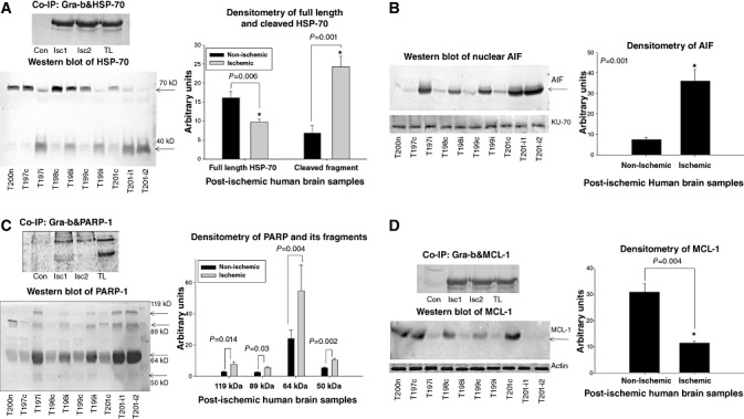
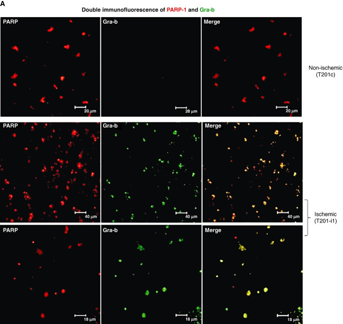
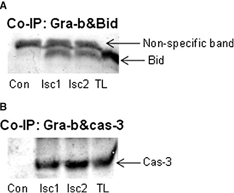
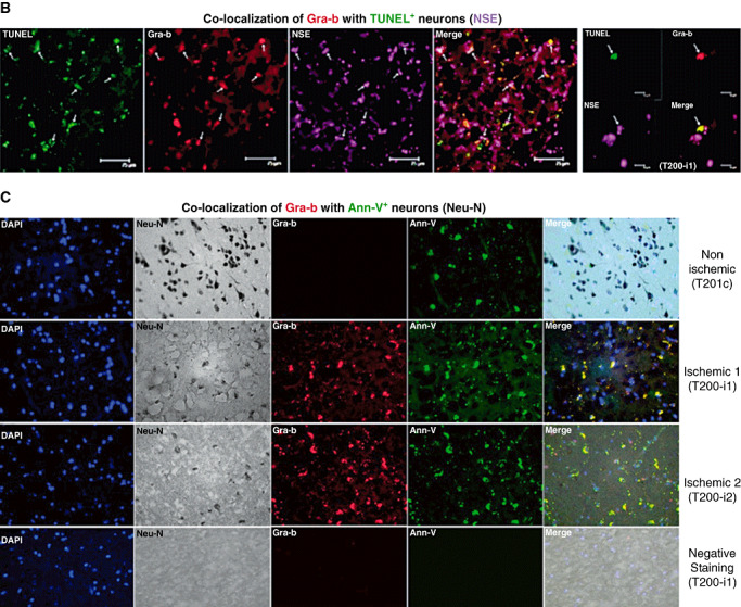
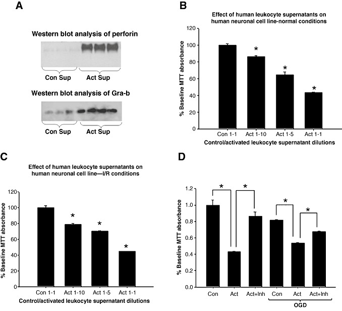
Infiltration of leukocytes into post-ischemic cerebrum is a well-described phenomenon in stroke injury. Because CD-8(+) T-lymphocytes secrete cytotoxic proteases, including granzyme-b (Gra-b) that exacerbates post-ischemic brain damage, we investigated roles of Gra-b in human stroke. To study the role of Gra-b in stroke, ischemic and non-ischemic tissues (from post-mortem stroke patients) were analyzed using immunoblotting, co-immunoprecipitation, terminal deoxy uridine nick end labeling (TUNEL) and Annexin-V immunostaining, and in vitro neuron survival assays. Activated CG-SH cells and supernatants were used to model leukocyte-dependent injury. Non-ischemic brain tissues were used as non-pathological controls. Non-activated CG-SH cells and supernatants were used as controls for in vitro experiments. Human stroke (ischemic) samples contained significantly higher levels of Gra-b and interferon-gamma inducible protein-10 (IP-10/CXCL10) than non-ischemic controls. In stroke, poly (ADP-ribose) polymerase-1 and heat shock protein-70 were cleaved to canonical proteolytic "signature" fragments by Gra-b. Gra-b was also found to bind to Bid and caspase-3. Gra-b also co-localized with Annexin-V(+) /TUNEL(+) in degenerating neurons. Importantly, Gra-b inhibition protected both normal and ischemia-reperfused neurons against in vitro neurotoxicity mediated by activated CG-SH cells and supernatants. These results suggest that increased leukocyte infiltration and elevated Gra-b levels in the post-stroke brain can induce contact-dependent and independent post-ischemic neuronal death to aggravate stroke injury.


Conflict of interest statement

The authors declare no conflict of interest.

Figures

Figure 1
Figure 1
Hematoxylin and eosin staining shows increased apoptosis and necrosis in the post‐mortem human stroke tissue. Human post‐mortem stroke samples were processed and stained with hematoxylin and eosin. Tissues obtained are from the infarct zone and away from the infarct (considered non‐ischemic tissue) which increased the homogeneity of the tissue to be used as control for the ischemic tissue. Deranged brain tissue with several degenerating cells (pointed with small arrows and arrow heads), shrunken nucleus, disrupted neuropil and extensive disruption of brain parenchyma and matrix (pointed with big arrows) is seen in the ischemic tissue. Moreover, most of the ischemic samples were heavily stained with eosin indicating the edematous condition of the tissue (indicated with *). Increased eosin staining was observed in all the ischemic samples over controls. Slight derangement of tissue was also observed occasionally in the control tissue but essentially looked similar to normal. Normal (non‐ischemic) brain tissue from patient sample ID 08/HBTR/T200 was obtained from cortical region. The regions and types of tissues obtained were outlined in the Table 1.
Figure 2
Figure 2
A.Post‐mortem human stroke tissue contains elevated IP‐10/CXCL10 and Gra‐b levels. Immunoblot analysis of IP‐10/CXCL10 shows a significant increase in IP‐10/CXCL10 levels in the ischemic human stroke samples (ipsilateral) over the non‐ischemic controls (contralateral). B. Immunoblot analysis of Gra‐b also shows a significant increase in the Gra‐b levels in all ischemic human stroke samples (ipsilateral) over the non‐ischemic samples (contralateral). T200n‐normal brain tissue; T197c, T97i‐contralateral and ipsilateral tissue from HBTR‐197; T198c, T198i‐contralateral and ipsilateral tissue from HBTR‐198; T199c, T199i‐contralateral and ipsilateral tissue from HBTR‐199; T201c, T201‐i1, T201‐i2‐contralateral and ipsilateral tissue from HBTR‐201. Unpaired t‐test with two‐tail P value, bars standard error (SE) * denotes significance.
Figure 3
Figure 3
Human ischemic infarcts contain increased numbers of Gra‐b+ CD‐8+ T‐cells. Double immunofluorescence analysis of CD‐8 and Gra‐b showed increased numbers of CD‐8+ T cells (FITC fluorescence‐ green) that were also Gra‐b+ (Cy3 fluorescence‐red) in human ischemic post‐mortem stroke samples compared with corresponding non‐ischemic controls. Representative immunofluorescent images are from the patient's samples with IDs’ T201c, T201‐i1.
Figure 4
Figure 4
A. Gra‐b proteolyzes HSP‐70, PARP‐1 and MCL‐1 and facilitates the nuclear translocation of AIF. Co‐immunoprecipitation (co‐IP) of Gra‐b with HSP‐70. Increased binding of Gra‐b with HSP‐70 can be observed. Representative image is from the “T201” which has two separate regions of the ischemic infarct. Immunoblot of HSP‐70. Significantly decreased level of HSP‐70 correlated with increased 40 kD proteolytic signature fragment mediated by Gra‐b can be observed. B. Immunoblot of AIF on nuclear samples. A significant increase in the levels of nuclear AIF can be observed. KU‐70 was used as a loading control for nuclear samples. C. Co‐IP of Gra‐b with PARP‐1. Increased binding of Gra‐b with PARP‐1 can be observed (119 kD). Immunoblot of PARP‐1. Significant increase in the cleaved signature fragments 89 and 64 kD can be observed. Further faint increase in the levels of 50 kD cathepsin‐b and can also be observed. D. Co‐IP of Gra‐b with MCL‐1. Increased binding of Gra‐b with MCL‐1 can be observed. Immunoblot of MCL‐1. Significant decrease in the levels of MCL‐1 in all the ischemic samples over the contralateral was observed. Representative image for Co‐IP is from the “T201” which has two non‐overlapping regions of the ischemic infarct. Unpaired t‐test with two tailed P value was obtained between two specific groups. Con‐ Non ischemic tissue, Isc1, Isc2‐ Ischemic tissue lysates, TL‐ Non immunoprecipitated ischemic tissue lysate (used as positive control).
Figure 5
Figure 5
A. Gra‐b co‐localizes with PARP‐1 and HSP‐70 in the degenerating cells of human post‐mortem ischemic infarct samples. Double immunofluorescence of Gra‐b and PARP‐1. Increased immunoreactivity for cells stained positive with both Gra‐b and PARP‐1 can be observed in ischemic samples over the contralateral samples. B. Double immunofluorescence of Gra‐b and HSP‐70. Increased immunoreactivity for cells stained positive with both Gra‐b and HSP‐70 can be observed in ischemic samples over the contralateral. Unpaired test with two‐tail P value, bars SE. Representative immunofluorescent images are from the patient's samples with IDs’ T201c, T201‐i1.
Figure 5
Figure 5
A. Gra‐b co‐localizes with PARP‐1 and HSP‐70 in the degenerating cells of human post‐mortem ischemic infarct samples. Double immunofluorescence of Gra‐b and PARP‐1. Increased immunoreactivity for cells stained positive with both Gra‐b and PARP‐1 can be observed in ischemic samples over the contralateral samples. B. Double immunofluorescence of Gra‐b and HSP‐70. Increased immunoreactivity for cells stained positive with both Gra‐b and HSP‐70 can be observed in ischemic samples over the contralateral. Unpaired test with two‐tail P value, bars SE. Representative immunofluorescent images are from the patient's samples with IDs’ T201c, T201‐i1.
Figure 6
Figure 6
A. Gra‐b interacts with pro‐apoptotic Bid and caspase‐3 in the human post‐mortem ischemic infarcts. Co‐immunoprecipitation (co‐IP) of Gra‐b with Bid. Increased binding of Gra‐b with Bid can be clearly observed. B. Co‐IP of Gra‐b with caspase‐3. Increased binding of Gra‐b with caspase‐3 can be observed. Representative image for Co‐IP is from the “T201” which has two non‐overlapping regions of the ischemic infarct. Con‐ Non ischemic tissue, Isc1, Isc2‐ Ischemic tissue lysates, TL‐ Non immunoprecipitated ischemic tissue lysate (used as positive control).
Figure 7
Figure 7
A. Apoptotic neurons in the human post‐mortem ischemic infarcts contain higher levels of translocated Gra‐b. Double immunofluorescence analysis of Gra‐b and MAP‐2. Increased presence of translocated Gra‐b (Cy3 fluorescence‐red) in the neurons stained positive for MAP‐2 (FITC fluorescence‐green) can be observed in the ischemic samples over the contralateral. Most of the cells positive for MAP‐2 appeared to be degenerating because of their shrunken shape, lost of cellular structure and disorganized neuropil. Representative immunofluorescent images are from the patient's samples with IDs’ T201c, T201‐i1. B. Double immunofluorescence analysis of Gra‐b and NSE with TUNEL. Increased number of cells positive for TUNEL (FITC fluorescence‐green), Gra‐b (Cy3 fluorescence‐red) and NSE (Cy5 fluorescence‐violet) can be observed in the ischemic samples. Representative immunofluorescent image is from the patient's sample with ID T200‐i1. C. Triple immunostaining of Gra‐b, Neu‐N and Annexin‐V (Ann‐V). Increased immunoreactivity of cells for Gra‐b (Cy3 fluorescence‐red), Neu‐N DAB stained and Ann‐V (FITC fluorescence‐green) can be observed in the human ischemic tissue compared with non‐ischemic human stroke samples. No immunoreactivity is observed in negative staining. Representative immunofluorescent images are from the patient's samples with IDs’ T201c, T201‐i1, T201‐i2.
Figure 7
Figure 7
A. Apoptotic neurons in the human post‐mortem ischemic infarcts contain higher levels of translocated Gra‐b. Double immunofluorescence analysis of Gra‐b and MAP‐2. Increased presence of translocated Gra‐b (Cy3 fluorescence‐red) in the neurons stained positive for MAP‐2 (FITC fluorescence‐green) can be observed in the ischemic samples over the contralateral. Most of the cells positive for MAP‐2 appeared to be degenerating because of their shrunken shape, lost of cellular structure and disorganized neuropil. Representative immunofluorescent images are from the patient's samples with IDs’ T201c, T201‐i1. B. Double immunofluorescence analysis of Gra‐b and NSE with TUNEL. Increased number of cells positive for TUNEL (FITC fluorescence‐green), Gra‐b (Cy3 fluorescence‐red) and NSE (Cy5 fluorescence‐violet) can be observed in the ischemic samples. Representative immunofluorescent image is from the patient's sample with ID T200‐i1. C. Triple immunostaining of Gra‐b, Neu‐N and Annexin‐V (Ann‐V). Increased immunoreactivity of cells for Gra‐b (Cy3 fluorescence‐red), Neu‐N DAB stained and Ann‐V (FITC fluorescence‐green) can be observed in the human ischemic tissue compared with non‐ischemic human stroke samples. No immunoreactivity is observed in negative staining. Representative immunofluorescent images are from the patient's samples with IDs’ T201c, T201‐i1, T201‐i2.
Figure 8
Figure 8
A. Activated CG‐SH cells secrete increased levels of Gra‐b and perforin and can induce Gra‐b dependent neurotoxicity. Immunoblots of perforin and Gra‐b. Immunoblot of non‐activated and activated CG‐SH supernatants showed higher levels of Gra‐b and perforin in activated CG‐SH supernatants than non‐activated controls. B. Activated CG‐SH cell supernatants induce neurotoxicity in human SH‐SY5Y neuronal cells in normal conditions in a dose dependent fashion compared with controls. MTT was significantly and dose dependently decreased with activated CG‐SH cell supernatants than non‐activated CG‐SH cell supernatants. C. Activated CG‐SH also induced neurotoxicity in ischemia challenged human SH‐SY5Y neurons in a dose dependent fashion assessed by decrease in MTT conversion. D. Gra‐b inhibition (50 µM) protected normal and ischemia challenged human SH‐SY5Y neurons from activated CG‐SH cells (leukocyte model) induced neurotoxicity mediated via Gra‐b.

Similar articles

Cited by

References

    1. Sherman DG, Bes A, Easton JD, Hacke W, Kaste M, Polmar SH et al (2001) Use of anti‐ICAM‐1 therapy in ischemic stroke: results of the Enlimomab Acute Stroke Trial. Neurology 57:1428–1434. - PubMed
    1. Alexander JS, Dayton T, Davis C, Hill S, Jackson TH, Blaschuk O et al (1998) Activated T‐lymphocytes express occludin, a component of tight junctions. Inflammation 22:573–582. - PubMed
    1. Alimonti JB, Shi L, Baijal PK, Greenberg AH (2001) Granzyme B induces BID‐mediated cytochrome c release and mitochondrial permeability transition. J Biol Chem 276:6974–6982. - PubMed
    1. Amantea D, Nappi G, Bernardi G, Bagetta G, Corasaniti MT (2009) Post‐ischemic brain damage: pathophysiology and role of inflammatory mediators. FEBS J 276:13–26. - PubMed
    1. Andersen MH, Becker JC, Thor SP (2005) The antiapoptotic member of the Bcl‐2 family Mcl‐1 is a CTL target in cancer patients. Leukemia 19:484–485. - PubMed

Publication types

MeSH terms