. 2010 Oct;101(10):2269-77.
doi: 10.1111/j.1349-7006.2010.01674.x. Epub 2010 Sep 1.

Expression of C4.4A at the invasive front is a novel prognostic marker for disease recurrence of colorectal cancer

Affiliations

Expression of C4.4A at the invasive front is a novel prognostic marker for disease recurrence of colorectal cancer

Ken Konishi et al. Cancer Sci. 2010 Oct.

Erratum in

  • Cancer Sci. 2010 Nov;101(11):2503

Abstract

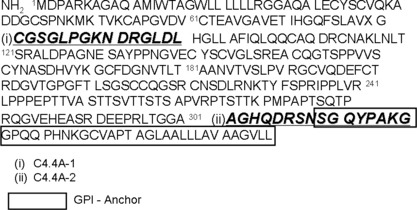
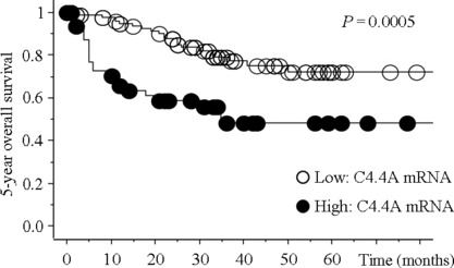
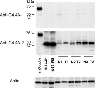
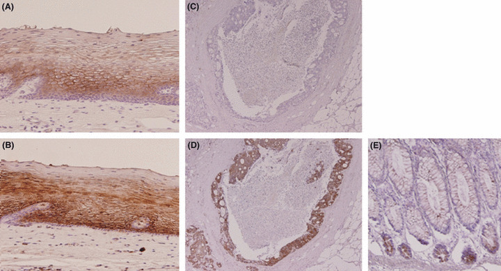
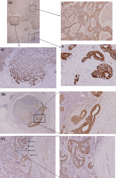
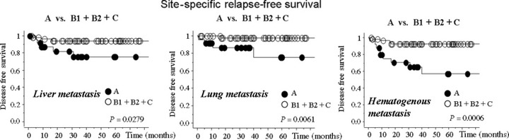
Metastasis-associated gene C4.4A is a glycolipid-anchored membrane protein expressed in several human malignancies. The aim of this study was to explore the expression and clinical relevance of C4.4A in colorectal cancer. By quantitative RT-PCR, 154 colorectal cancer tissues were examined for C4.4A mRNA. We examined 132 colorectal cancer tissues by immunohistochemistry using a new polyclonal antibody that recognizes the C4.4A protein C-terminus containing the glycosylphosphatidyl-inositol anchor signaling sequence. A significant difference in 5-year overall survival was found between samples with high and low expression of C4.4A mRNA (P = 0.0005). Immunohistochemistry showed strong membranous staining of C4.4A at the invasive front of colorectal cancer tumors and at the frontier of metastatic lesions to lymph node and lung. The membranous staining with enhanced intensity at the invasive front of the primary colorectal cancer (Type A: 34/132, 25.6%) was associated with depth of invasion (P = 0.033) and venous invasion (P = 0.003), and was a significant independent prognostic factor (5-year overall survival in the entire series [n = 132; P = 0.004] and disease-free survival in stage II and III colorectal cancers [n = 82; P = 0.003]). Moreover, Type A C4.4A expression was linked to shorter liver metastasis-free survival rate, lung metastasis-free survival rate, or hematogenous metastasis-free survival (P = 0.0279, P = 0.0061, and P = 0.0006, respectively). Our data indicate that expression of the C4.4A protein at the invasive front acts as a novel prognostic marker in colorectal cancer, possibly through invasion-related mechanisms.


Figures

Figure 1
Figure 1
Amino acid sequences of C4.4A protein and epitopes used as immunogens. (i) CGSGLPGKNDRGLDL near the N‐terminus, and (ii) AGHQDRSNSGQYPAKG at the C‐terminus. The boxed area indicates the glycosylphosphatidyl‐inositol (GPI) anchor signaling sequence.
Figure 2
Figure 2
Kaplan–Meier plots showing survival curves stratified by C4.4A mRNA expression in colorectal cancer tissues. A total of 154 colorectal cancer tissues were examined for C4.4A mRNA expression using quantitative RT‐PCR. A significant difference in 5‐year overall survival rate was noted between high and low C4.4A expression (P = 0.0005).
Figure 3
Figure 3
Western blot analysis of the C4.4A protein in normal esophageal tissue, colorectal cancer cell lines (HCT116 and KM12SM), and in three samples from patients taken from normal (N) and cancerous colorectal tissues (T). Esophageal epithelium displayed an intense band of approximately 67 kDa with both the anti‐C4.4A‐1 and anti‐C4.4A‐2 antibodies. However, in both colon cancer cell lines and colon tissue samples, anti‐C4.4A‐2 antibody alone displayed a prominent band at approximately 40 kDa. Anti‐C4.4A‐1 antibody did not produce any bands. The blot for actin served as a loading control.
Figure 4
Figure 4
Tissue sections of normal esophageal mucosa (A,B) and colorectal cancer tissue (C,D) were immunostained with anti‐C4.4A‐1 (A,C) and anti‐C4.4A‐2 (B,D) antibodies. (A,B) Membranous staining was observed in the suprabasal squamous epithelium. (C) No signal was observed with the C4.4A‐1 antibody in a colorectal cancer tissue sample. (D) Strong staining was observed with anti‐C4.4A‐2 antibody on the same sample. (E) Tissue sections of normal colonic mucosa. In normal colonic mucosa, staining was observed with anti‐C4.4A‐2 antibody at the bottom of the gland. Magnification, (A,B) ×100; (C–E) ×40.
Figure 5
Figure 5
Immunohistochemistry of C4.4A expression in colorectal cancer. (A) Unique expression pattern of C4.4A at the invasive front of specimens from colorectal cancer. (A‐I) In the superficial or intermediate portion, weak C4.4A expression was observed mostly in the cytoplasm. (A‐II) C4.4A expression was present on the plasma membrane at the invasive front. (A‐III) Among the expansive population of cancer specimens, only those cells that faced virgin stroma expressed the C4.4A protein. (B) Metastatic lesion to lymph node. Metastatic tumor cells invading into the fibrous capsule exclusively expressed intense, membranous C4.4A. (C) Metastatic lesion from the lung. Metastatic tumor cells displayed a similar expression pattern of C4.4A at the invasive margin (indicated by arrows). Magnification, (A) upper left, ×12.5; (A‐I,II) ×100; (A‐III) ×40; (B) left, ×12.5; right, ×100; (C) left, ×40; right, ×100.
Figure 6
Figure 6
Staining pattern of C4.4A and prognosis of patients with colorectal cancer. Kaplan–Meier 5‐year overall survival (OS) curves for patients with stage I–IV tumors (n = 132). The cases were categorized as: (A) translocation from cytoplasm to membrane and with enhanced intensity (n = 34); (B1) translocation but without enhanced intensity (n = 12); (B2) no translocation but with enhanced intensity (n = 15); or (C) no translocation and without enhanced intensity (n = 71). Change of localization from cytoplasm to the plasma membrane (A + B1) or increase in intensity (A + B2) at the invasive front was associated with shorter 5‐year OS (P = 0.023 and P = 0.0031, respectively). More striking differences were noted between Type A and other expression and localization types (B1 + B2 + C) (P = 0.0013). (B) The 5‐year disease‐free survival in patients with stage II and III tumors (n = 82). When these patients were analyzed for 5‐year disease‐free survival after curative surgery, similar results to those in 5‐year OS were obtained (P = 0.011, P = 0.0016, and P = 0.0004, respectively).
Figure 7
Figure 7
Site‐specific relapse‐free survival in colorectal cancer patients with stage II and III tumors (n = 82). The patients in stage II and III developed disease recurrence: liver metastasis (n = 8); lung metastasis (n = 5); bone metastasis (n = 1); local recurrence (n = 3); and lymph node metastasis (n = 3). Among them, one patient had both liver metastasis and lung metastasis, and another had both liver metastasis and local recurrence. The cases were categorized as: (A) translocation from cytoplasm to membrane and with enhanced intensity; (B1) translocation but without enhanced intensity; (B2) no translocation but with enhanced intensity; or (C) no translocation and without enhanced intensity. Type A C4.4A expression was linked to shorter liver metastasis‐free survival rate, lung metastasis‐free survival rate, or hematogenous metastasis‐free survival (P = 0.0279, P = 0.0061, and P = 0.0006, respectively). No difference was observed in other site‐specific disease‐free survival curves (data not shown).
Figure 8
Figure 8
In vitro cultures of HCT116 colon cancer cells. (A) The C4.4A protein expression was observed in the nucleus of HCT116 in 2D culture. (B) The 3D cell cultures in collagen gels. The HCT116 cells on the gels expressed C4.4A protein in the cytoplasm, whereas the protein was often localized to the plasma membrane when the cells were surrounded by a collagen matrix. Note that the collagen matrix tended to become degraded when the cells had membranous C4.4A expression. The arrows indicate intense membranous staining. Magnification, ×200.

Similar articles

Cited by

References

    1. National Cancer Institute . Colon and rectal cancers. [Cited July 1, 2009.] Available from URL: http://www.cancer.gov/cancertopics/types/colon‐and‐rectal.
    1. Wadhawan A, Stephens R, Adams R. Intermittent therapy in the palliative treatment of metastatic colorectal cancer. Expert Rev Anticancer Ther 2009; 9: 125–34. - PubMed
    1. Hurwitz H, Fehrenbacher L, Novotny W et al. Bevacizumab plus irinotecan, fluorouracil, and leucovorin for metastatic colorectal cancer. N Engl J Med 2004; 350: 2335–42. - PubMed
    1. Van Cutsem E, Köhne CH, Hitre E et al. Cetuximab and chemotherapy as initial treatment for metastatic colorectal cancer. N Engl J Med 2009; 360: 1408–17. - PubMed
    1. Matzku S, Wenzel A, Liu S, Zöller M. Antigenic differences between metastatic and nonmetastatic BSp73 rat tumor variants characterized by monoclonal antibodies. Cancer Res 1989; 49: 1294–9. - PubMed

Publication types