. 2010 Dec;139(6):2072-2082.e5.
doi: 10.1053/j.gastro.2010.08.053. Epub 2010 Oct 19.

Characterization of the intestinal cancer stem cell marker CD166 in the human and mouse gastrointestinal tract

Affiliations

Characterization of the intestinal cancer stem cell marker CD166 in the human and mouse gastrointestinal tract

Trevor G Levin et al. Gastroenterology. 2010 Dec.

Abstract

Background & aims: CD166 (also called activated leukocyte cell adhesion molecule [ALCAM]) is a marker of colorectal cancer (CRC) stem cells; it is expressed by aggressive tumors. Although the presence of CD166 at the tumor cell surface has been correlated with shortened survival, little is known about its function and expression in normal intestinal epithelia.

Methods: We characterized the expression pattern of CD166 in normal intestinal tissue samples from humans and mice using immunohistochemisty, flow cytometry, and quantitative reverse-transcriptase polymerase chain reaction. Human and mouse intestinal tumors were also analyzed.

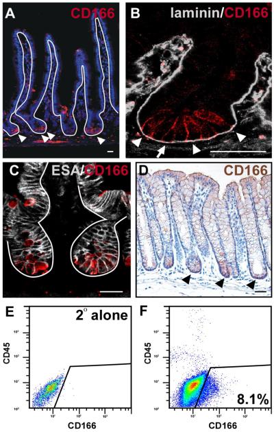
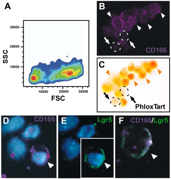
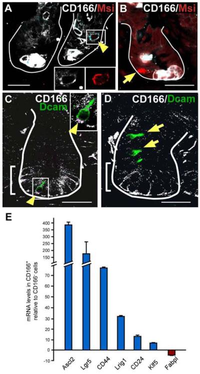
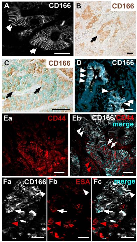
Results: CD166 was expressed on the surface of epithelial cells within the stem cell niche and along the length of the intestine; expression was conserved across species. In the small intestine, CD166 was observed on crypt-based Paneth cells and intervening crypt-based columnar cells (putative stem cells). A subset of CD166-positive, crypt-based columnar cells coexpressed the stem cell markers Lgr5, Musashi-1, or Dcamkl-1. CD166 was located in the cytoplasm and at the surface of cells within human CRC tumors. CD166-positive cells were also detected in benign adenomas in mice; rare cells coexpressed CD166 and CD44 or epithelial-specific antigen.

Conclusions: CD166 is highly expressed within the endogenous intestinal stem cell niche. CD166-positive cells appear at multiple stages of intestinal carcinoma progression, including benign and metastatic tumors. Further studies should investigate the function of CD166 in stem cells and the stem cell niche, which might have implications for normal intestinal homeostasis. CD166 has potential as a therapeutic target for CRC.


Figures

Figure 1
Figure 1. CD166 expression pattern in the human small intestine and colon
(A-C) Human small intestine stained with antibodies to CD166 (red) and counterstained with Hoechst (blue). (A) CD166-positive cells are located within the mesenchymal (arrows) and epithelial compartments (arrowheads). (Ba-Bb) Enlarged view of small intestinal crypts. (C-Da,Db) Human colon stained with antibodies to CD166, visualized with DAB (brown) in (C) and with fluorescence (red) and Hoechst counterstain (blue) in (Da-Db). Arrowheads point to cell surface epithelial expression. (E,F) larger magnification views of the crypt base boxed in yellow from B and D, respectively. Solid lines demark epithelial-mesenchymal boundary and dashed lines mark the apical epithelial surface. Bar=25μm.
Figure 2
Figure 2. CD166 expression pattern in the mouse small intestine and colon
(A-B) Mouse small intestine (SI) stained with antibodies to CD166 (red) and (A) counterstained with Hoechst (blue) or (B) co-stained with antibodies to laminin (gray) in higher magnification of the crypt. Arrowheads mark CD166-expressing cells. Arrows mark columnar crypt-based cell in B. (C) Mouse small intestinal crypt co-stained with CD166 (red) and the pan-epithelial ESA (white). (D) Mouse colon stained with antibodies to CD166 (brown), demonstrating enhanced expression in the crypt base (black arrowheads). Solid lines denote the epithelial-mesenchymal boundary. Bar=25μm. (E) Flow cytometry isotype control on isolated intestinal epithelial crypt cells. (F) Flow cytometry analysis of isolated crypt epithelial cells stained with CD166 antibodies. Box denotes CD166-positive, CD45-negative cells.
Figure 3
Figure 3. FACS-isolated CD166 epithelial cells express Paneth cell granules or Lgr5
(A) Forward (FSC) and side (SSC) scatter analysis of CD166-expressing crypt epithelial cells display two distinct populations of cells (orange centers). (B-C) FACS-isolated, cytospun CD166-expressing mouse crypt cells (B; purple) stained with (C) Phloxine Tartrazine. White arrowheads designate CD166-positive cells; orange arrowheads designate CD166-positive, Phloxine Tartrazine-positive cells. Arrows and dashed circles designate CD166-positive, Phloxine Tartrazine-negative cells. (D-F) Cytospun, isolated CD166-positive cells (purple) co-stained with antibodies to Lgr5, a putative stem cell marker (green). Arrowhead designates a double-labeled cell.
Figure 4
Figure 4. CD166 is expressed in a subset of cells expressing stem cell markers Musashi-1 and Dcamkl-1
(A,B) Mouse small intestinal tissue co-labeled with antibodies to CD166 (white) and the putative stem cell marker, Musashi-1 (Msi, red). (A) White box and yellow arrowhead designates a CD166/Msi co-expressing cell with cell surface co-expressing in light blue. Bottom right insets depict higher magnification of singly stained cell, CD166 (white) and Msi (red). (B) A Msi-positive, CD166-negative cells indicated with arrow. (C,D) Mouse intestinal tissue co-labeled with antibodies to CD166 (white) and the putative stem cell marker, Dcamkl-1 (green). (C) Yellow arrowhead and box designates a CD166/Dcamkl co-expressing cell, with the co-expressing cell surface in purple. A higher magnification is provided in the upper right inset. (D) CD166-negative, Dcamkl-1-positive cells are designated by arrowhead. Brackets mark CD166-expressing region. White lines mark epithelial-mesenchymal boundary. Bar=25μm. (E) Quantitative RT-PCR analysis of stem cell markers within the CD166-positive cell population relative to the adjacent CD166-negative population. Triplicate samples of n=2 runs, and S.E.M.
Figure 5
Figure 5. CD166 expression of human colorectal cancer is recapitulated in mouse colorectal adenomas
(A-B) Human primary colorectal adenomas labeled with antibodies to CD166 show (A) cell surface expression (white, arrowheads) or (B) cytoplasmic expression (brown, arrow). (C) Human colorectal liver metastasis that has both cell surface and cytoplasmic expression of CD166 (brown, arrow). Methyl Green nuclear counterstain (green). (D) Benign mouse intestinal tumor labeled with antibodies to CD166 (white, arrowheads) and Hoechst nuclear counterstain (blue). (Ea-Eb) Human primary colorectal adenoma co-stained with antibodies to CD44 (red) and CD166 (white). (Eb) Subpopulations of tumor cells express both CD44 and CD166 (merged, light blue, white arrows), or only CD44 (red staining, red arrowheads). Some tumor cells do not express CD44 but express CD166 (white staining, white arrowheads). (Fa-Fc) Mouse adenoma stained with antibodies to CD166 (white) and ESA (red). Subpopulations of cells express CD166 and ESA (merged, light blue, white arrow), ESA alone (red arrowhead) or CD166 alone (white arrowhead). Bar=25μm.

References

    1. Cancer Facts & Figures 2008. American Cancer Society; Atlanta: 2008.
    1. Cho RW, Clarke MF. Recent advances in cancer stem cells. Curr Opin Genet Dev. 2008;18:48–53. - PubMed
    1. Dylla SJ, Beviglia L, Park IK, et al. Colorectal cancer stem cells are enriched in xenogeneic tumors following chemotherapy. PLoS ONE. 2008;3:e2428. - PMC - PubMed
    1. Pardal R, Clarke MF, Morrison SJ. Applying the principles of stem-cell biology to cancer. Nat Rev Cancer. 2003;3:895–902. - PubMed
    1. Nowell PC. The clonal evolution of tumor cell populations. Science. 1976;194:23–8. - PubMed

Publication types

MeSH terms