. 2010 Dec;139(6):2050-2060.e2.
doi: 10.1053/j.gastro.2010.08.052. Epub 2010 Oct 16.

Bone morphogenetic protein signaling regulates gastric epithelial cell development and proliferation in mice

Affiliations

Bone morphogenetic protein signaling regulates gastric epithelial cell development and proliferation in mice

Masahiko Shinohara et al. Gastroenterology. 2010 Dec.

Abstract

Background & aims: We investigated the role of bone morphogenetic protein (BMP) signaling in the regulation of gastric epithelial cell growth and differentiation by generating transgenic mice that express the BMP inhibitor noggin in the stomach.

Methods: The promoter of the mouse H+/K+-ATPase β-subunit gene, which is specifically expressed in parietal cells, was used to regulate expression of noggin in the gastric epithelium of mice. The transgenic mice were analyzed for noggin expression, tissue morphology, cellular composition of the gastric mucosa, gastric acid content, and plasma levels of gastrin. Tissues were analyzed by immunohistochemical, quantitative real-time polymerase chain reaction, immunoblot, microtitration, and radioimmunoassay analyses.

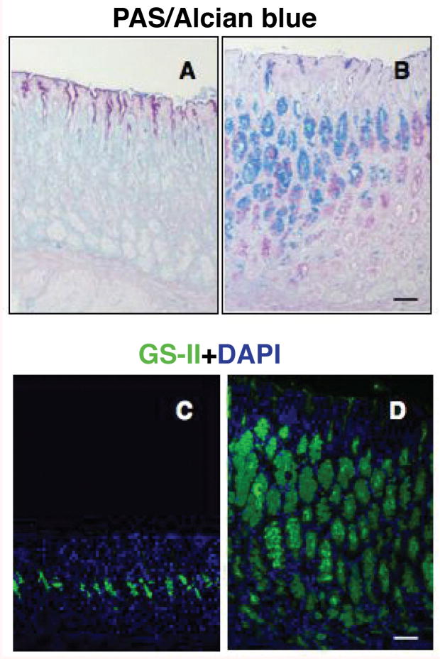
Results: In the stomachs of the transgenic mice, phosphorylation of Smad 1, 5, and 8 decreased, indicating inhibition of BMP signaling. Mucosa were of increased height, with dilated glands, cystic structures, reduced numbers of parietal cells, and increased numbers of cells that coexpressed intrinsic factor, trefoil factor 2, and Griffonia (Bandeiraea) simplicifolia lectin II, compared with wild-type mice. In the transgenic mice, levels of the H+/K+-ATPase α-subunit protein and messenger RNA were reduced, whereas those of intrinsic factor increased. The transgenic mice were hypochloridric and had an increased number of Ki67- and proliferating cell nuclear antigen-positive cells; increased levels of plasma gastrin; increased expression of transforming growth factor-α, amphiregulin, and gastrin; and activation of extracellular signal-regulated kinase 2.

Conclusions: Inhibiting BMP signaling in the stomachs of mice by expression of noggin causes loss of parietal cells, development of transitional cells that express markers of mucus neck and zymogenic lineages, and activation of proliferation. BMPs are therefore important regulators of gastric epithelial cell homeostasis.


Conflict of interest statement

Conflicts of interest The authors disclose no conflicts.

Figures

Figure 1
Figure 1
Generation of noggin TG mice. (A) Diagram of the H/K-Noggin transgene. Noggin was placed under the control of the mouse H+,K+-ATPase β-subunit promoter. A mutated human growth hormone (mut hGH) cassette provided intron and polyadenylation sequences. (B) Transgene messenger RNA (mRNA) levels in 12-week-old mice from TG-lines Nog.3 and Nog.4 were compared with background levels in non-TG mice using QRT-PCR and displayed as fold increase over the non-TG negative controls. Values are shown as means ± standard error, n = 4. (C) Gastric paraffin sections from 12-week-old non-TG and TG mice were stained with the anti-p-Smad1-5-8 antibody and a biotin-conjugated secondary antibody. Scale bars, 50 μm. Arrows point to p-Smad1-5-8 positive cells. The magnified window depicts rare p-Smad1-5-8 positive nuclei in epithelial cells of the TG mice. Similar results were observed in 3 other 12-week-old non-TG and TG mice. (D) Expression of p-Smad1, 5, and 8 and GAPDH in gastric extracts obtained from 12-week-old non-TG and TG mice from line Nog.4 was studied by Western blots. (E) Bar graphs represent results obtained from densitometric analysis of the blots. P-Smad 1, 5, and 8 expression levels were normalized to GAPDH expression. Data are expressed as means ± standard error, n = 4. *P < .05.
Figure 2
Figure 2
Development of morphologic abnormalities in the gastric epithelium of noggin TG mice. Representative H&E-stained gastric paraffin sections of the corpus of 4-week-old non-TG (A) and TG mice (B) and of 12-week-old non-TG (C) and TG mice (D and E). Scale bar, 50 μm. Similar histologic changes were observed in three 4-week-old and in seven 12-week-old TG mice from line Nog.4 and in four 12-week-old TG mice from line Nog.3.
Figure 3
Figure 3
Diminished parietal cell number in noggin TG mice. (A) Number of cells per gland exhibiting the morphologic appearance of parietal cells in H&E-stained sections from 12-week-old TG and non-TG mice. Values are shown as means ± standard error, n = 3. *P < .01. The sections from 12-week-old mice were stained with the anti-H+,K+-ATPase α-subunit antibody and a FITC-conjugated secondary antibody (B). Scale bar, 50 μm. Similar results were observed in 7 other 12-week-old TG mice. H+,K+-ATPase α-subunit protein expression in non-TG and TG mice was measured by Western blots (C). Basal gastric acid content in non-TG and TG mice was determined by automated titration (C). Acid content values are shown as means ± standard error, n = 3. *P < .05.
Figure 4
Figure 4
Expression of Alcian blue-positive mucus and expansion of mucous neck cells in noggin TG mice. Gastric paraffin sections from non-TG (A and C) and TG mice (B and D) were stained with periodic acid Schiff (PAS)/Alcian blue (top panels) and Alexa 488-GS II (bottom panels). Scale bar, 50 μm. Similar results were observed in 3 other 12-week-old TG mice.
Figure 5
Figure 5
Expression of noggin in TG mice leads to increased IF-expressing cells. Gastric paraffin sections from non-TG and TG mice were stained with the IF antibody and a biotin-conjugated secondary antibody (A). Scale bar, 50 μm. Similar results were observed in 7 other 12-week old TG mice. IF messenger RNA (mRNA) abundance in non-TG mice was compared with that detected in TG mice using QRT-PCR and displayed as fold increase over the non-TG negative controls (B). Values are shown as means ± standard error, n = 8. *P < .05. IF protein levels in non-TG and TG mice were measured by Western blots with anti-IF antibodies (C).
Figure 6
Figure 6
Alterations in gastric epithelial cell differentiation in noggin TG mice. Gastric paraffin sections from non-TG and TG mice were stained with anti-IF primary antibodies and Cy-3 conjugated secondary antibodies (red) together with Alexa 488-conjugated GS II (green) (A and B), anti-TFF2 primary antibodies, and Cy-3-conjugated secondary antibodies (red) together with anti-IF primary antibodies and FITC-conjugated secondary antibodies (green) (C and D). Scale bar, 50 μm. Arrows point to cells expressing both markers. Similar results were observed in 2 other TG mice. QRT-PCR analysis of MUC-6 (E) and TFF2 (F) mRNA abundance in fundic RNA samples. Values are shown as fold change (means ± standard error, n = 4 for MUC-6 and n = 8 for TFF2) compared with non-TG levels. *P < .05.
Figure 7
Figure 7
Increased cell proliferation and expression of mitogenic signaling pathways in noggin TG mice. (A) Gastric paraffin sections from non-TG and TG mice were stained with anti-proliferating cell nuclear antigen (PCNA) (top panels) and anti-Ki67 antibodies (bottom panels). Scale bar, 50 μm. (B) Graph bars represent the number of Ki67 and PCNA-positive nuclei detected in both TG- and non-TG littermates. Values are shown as means ± standard error, n = 4 for PCNA and n = 3 for Ki67. *P < .05. (C) TGF-α and amphiregulin mRNA signals in non-TG mice were compared with those detected in TG mice using QRT-PCR and displayed as fold increase over the non-TG negative controls. Values are shown as means ± standard error, n = 4. *P < .05. (D) Phosphorylation and activation of ERK2 in non-TG and TG mice was studied by Western blots using an anti-phospho-ERK2 antibody. Total ERK2 levels were monitored by Western blots with an antibody recognizing ERK2 independent of its phosphorylation state.

Comment in

References

    1. Winnier G, Blessing M, Labosky PA, et al. Bone morphogenetic protein-4 is required for mesoderm formation and patterning in the mouse. Genes Dev. 1995;9:2105–2116. - PubMed
    1. Dunn NR, Winnier GE, Hargett LK, et al. Haploinsufficient phenotypes in Bmp4 heterozygous null mice and modification by mutations in Gli3 and Alx4. Dev Biol. 1997;188:235–247. - PubMed
    1. Kawabata M, Imamura T, Miyazono K. Signal transduction by bone morphogenetic proteins. Cytokine Growth Factor Rev. 1998;9:49–61. - PubMed
    1. van den Brink GR, Hardwick JC, Tytgat GN, et al. Sonic hedgehog regulates gastric gland morphogenesis in man and mouse. Gastroenterology. 2001;121:317–328. - PubMed
    1. Madison BB, Braunstein K, Kuizon E, et al. Epithelial hedgehog signals pattern the intestinal crypt-villus axis. Development. 2005;132:279–289. - PubMed

Publication types

MeSH terms