. 2011 Jan 28;51(2):269-79.
doi: 10.1016/j.visres.2010.08.039. Epub 2010 Sep 6.

Morphologies of mouse retinal ganglion cells expressing transcription factors Brn3a, Brn3b, and Brn3c: analysis of wild type and mutant cells using genetically-directed sparse labeling

Affiliations

Morphologies of mouse retinal ganglion cells expressing transcription factors Brn3a, Brn3b, and Brn3c: analysis of wild type and mutant cells using genetically-directed sparse labeling

Tudor Constantin Badea et al. Vision Res. .

Abstract

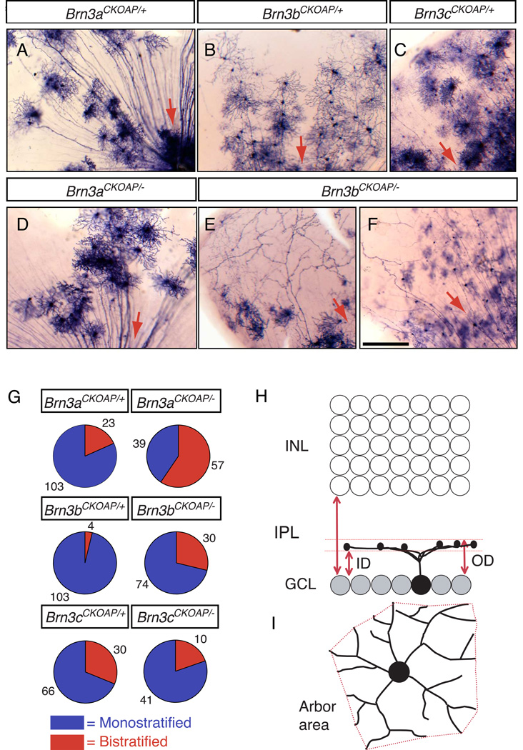
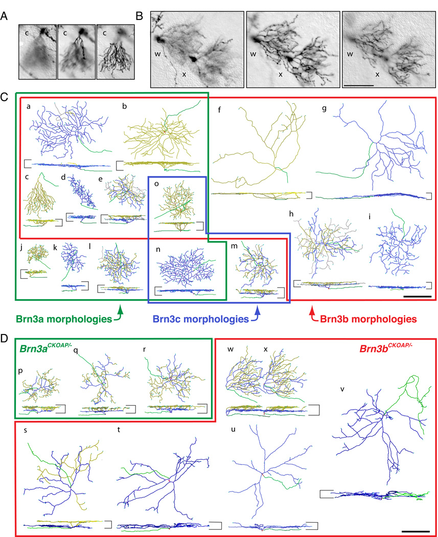
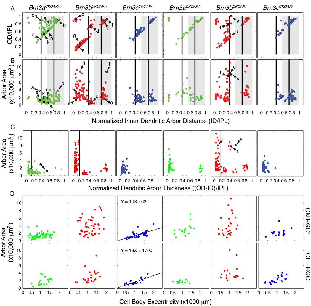
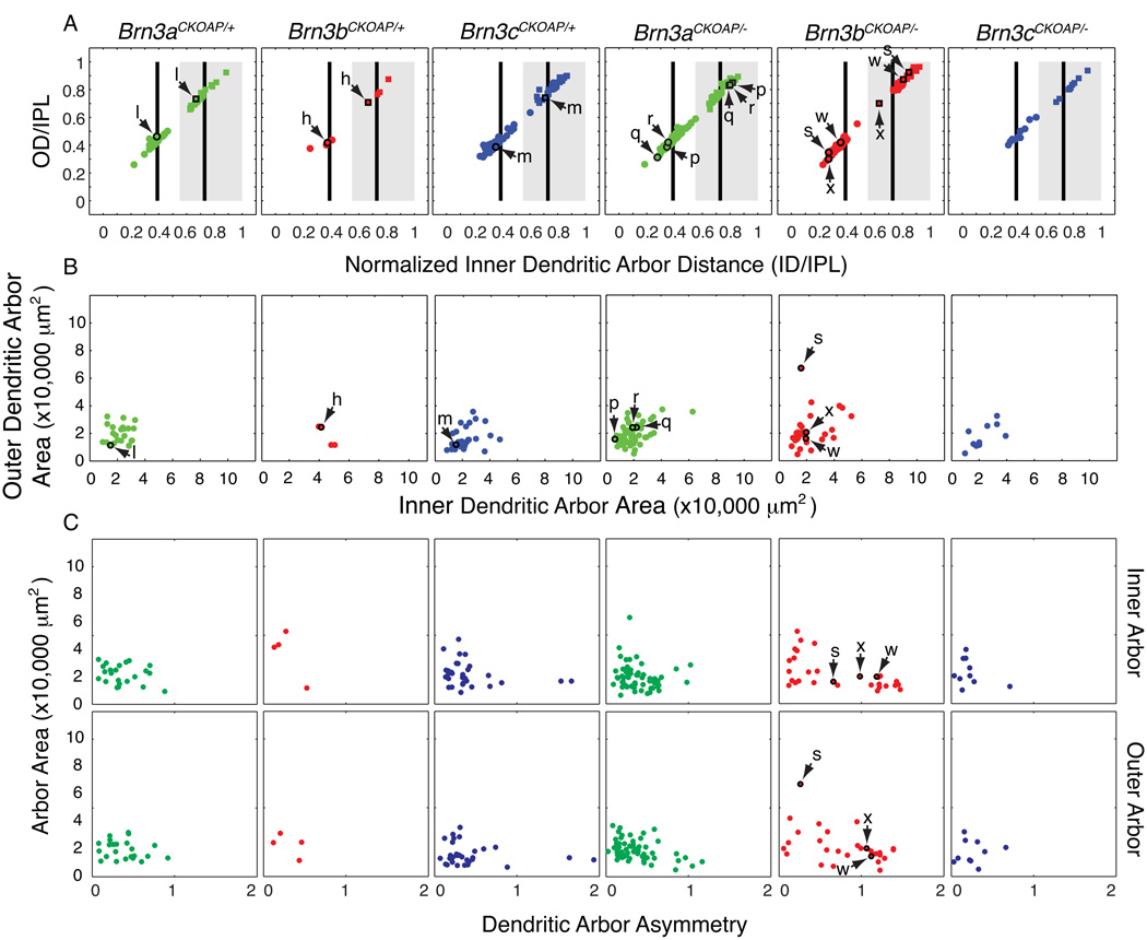
The mammalian retina contains more than 50 distinct neuronal types, which are broadly classified into several major classes: photoreceptor, bipolar, horizontal, amacrine, and ganglion cells. Although some of the developmental mechanisms involved in the differentiation of retinal ganglion cells (RGCs) are beginning to be understood, there is little information regarding the genetic and molecular determinants of the distinct morphologies of the 15-20 mammalian RGC cell types. Previous work has shown that the transcription factor Brn3b/Pou4f2 plays a major role in the development and survival of many RGCs. The roles of the closely related family members, Brn3a/Pou4f1 and Brn3c/Pou4f3 in RGC development are less clear. Using a genetically-directed method for sparse cell labeling and sparse conditional gene ablation in mice, we describe here the sets of RGC types in which each of the three Brn3/Pou4f transcription factors are expressed and the consequences of ablating these factors on the development of RGC morphologies.


Figures

Fig. 1
Fig. 1. Mouse RGCs visualized by sparse recombination of conditional AP reporter knock-in alleles at the Brn3a, Brn3b, and Brn3c loci
(A–F) Flat mount preparations of retinas from (A) R26rtTACreER/+;Brn3aCKOAP/+, (B) R26rtTACreER/+;Brn3bCKOAP/+, (C) R26rtT-CreER/+;Brn3cCKOAP/+, (D) R26rtTACreER/+;Brn3aCKOAP/−, and (E,F) R26rtTACreER/+;Brn3bCKOAP/− mice, histochemically stained for AP activity. Red arrows indicate the direction of the optic disc. (G) Pie charts showing the ratios of monostratified (blue): bistratified (red) RGCs from retinas of the indicated genotypes. The number of cells of each type is indicated. (H,I), Morphological parameters for the analysis of RGC dendritic arbors. In (H), the inner distance (ID) and outer distance (OD), normalized to the IPL thickness, together define the level of arbor stratification and thickness. ID and OD equal 0 at the level of the ganglion cell layer, and they equal 1 at the level of the INL. In (I), the arbor area is calculated as the area of the polygon (in red) with the shortest perimeter that connects the tips of the RGC dendrites when projected in the plane of the retina. Scale bar in F, 100 µm.
Fig. 2
Fig. 2. Examples of the morphologies of WT and mutant RGCs expressing each of the three Brn3AP alleles
(A,B) Sequential z-dimension DIC images of one Brn3bAP/+ (A) and two Brn3bAP/− (B) RGCs in flat mounted retinas. The planes of the optical sections are at the nerve fiber layer (left panels), at ~0.3–0.4 IPL depth (central panels) and at the boundary of the IPL and INL (right panels). The images in (A) and (B) correspond, respectively, to reconstructed cell c in panel (C) and reconstructed cells w and x in panel (D). For cell c, the dendrites stratify adjacent to the INL, whereas for cells w and x there are two distinct arbors (coded blue and yellow in (D)), one stratifying at an IPL depth of ~0.35 and the second stratifying adjacent to the INL. (C,D) Reconstructions of individual Brn3AP/+ (C) and Brn3AP/− (D) RGCs obtained from DIC z-stacks similar to the ones sampled in panels (A–B). For each cell, en face (top) and vertical views (bottom) are shown. The boundaries of the IPL are shown as horizontal bars on the sides of each vertical projection. Axons are green, vitreal (“ON”) dendrites are blue, and scleral (“OFF”) dendrites are yellow. “Recurrent” dendrites, defined as originating in the scleral plexus and ending in the vitreal plexus, are gray (present in cells a, e, h, l, and m). (C) Distinct RGC morphological types representative of the Brn3aAP/+, Brn3bAP/+ and Brn3cAP/+ data sets are enclosed in green, red and blue outlines, respectively. Types j, k, and l are unique to Brn3aAP/+ RGCs; types f, g, h and i to Brn3bAP/+ RGCs; and type m to Brn3cAP/+ RGCs. Types a–e are observed among both Brn3aAP/+ and Brn3bAP/+ RGCs; type n in Brn3aAP/+ and Brn3cAP/+ RGCs; and type o in all three Brn3AP/+ RGCs. The specific examples shown are derived from Brn3aCKOAP/+ (a, b, e, j, k, l, n), Brn3bCKOAP/+ (c, d, f, g, h, i, o) and Brn3cCKOAP/+ (m) retinas. (D) Examples of RGC morphologies observed in the Brn3aCKOAP/− and Brn3bCKOAP/− retinas are enclosed in green and red outlines, respectively. Examples of bistratified Brn3aAP/− RGCs (p, q, r), bistratified Brn3bAP/− RGCs (s, w, x) and monostratified Brn3bAP/− RGCs (t, u, v) are shown. Scale bars in B, C and D, 100 µm.
Fig. 3
Fig. 3. Morphological parameters for monostratified Brn3AP/+ and Brn3AP/− RGCs
Scatter plots in each vertical column refer to RGCs with the genotypes indicated at the top. Arrows and lower case letters identify the cells shown in Fig. 2. (A,B) Scatter plots of normalized inner distance (ID/IPL) vs. normalized outer distance (OD/IPL) and ID/IPL vs. dendritic arbor area. These parameters are defined in Fig. 1 H and I. The black vertical bars at 0.39 and 0.73 ID/IPL (referred to in the text as ~0.4 and ~0.7, respectively) represent the stratification levels of the “ON” (0.39) and “OFF” (0.73) bands of calretinin, calbindin, and choline acetyltransferase immunostaining, as measured by Haverkamp and Wassle (2000), Morgan et al. (2006), and Badea et al. (2009a). The grey rectangle spanning 0.55–1.0 ID/IPL represents the “OFF” sublamina of the IPL, as determined by the stratification level of the central band of calretinin/calbindin immunoreactivity (Haverkamp and Wassle, 2000; Badea et al 2009a) and of the axon arbor terminals of mGluR6-expressing, “ON” bipolar cells (Morgan et al 2006). (C) Scatter plots for the dendritic arbor thickness, (OD-ID)/IPL vs. arbor area. The dendritic arbor thickness represents the difference of the outer distance (OD) and inner distance (ID), as defined in Fig. 1H, normalized to the thickness of the IPL. (D) Dendritic arbor areas increase with eccentricity for flat monostratified Brn3cAP/+ and Brn3cAP/− RGCs in the mouse retina. Scatter plots of eccentricity (distance of the cell body from the optic nerve head) vs. area for flat monostratified RGCs. Flat monostratified RGCs include all monostratified Brn3cAP/+ RGCs, as well as Brn3aAP/+ and Brn3bAP/+ RGCs with normalized dendritic arbor thicknesses <0.12 and <0.15, respectively; these cutoffs are based on the positions of troughs in the arbor thickness distributions in panel C. For Brn3aAP/+ and Brn3bAP/+ RGCs, “ON” or “OFF” types were defined as having an ID/IPL ratio under or above 0.55, respectively. For Brn3cAP/+ RGCs, cells close to the 0.55 ON/OFF border were grouped with the RGCs that clustered nearby in parameter space in panels A and B. There is a linear correlation between eccentricity and dendritic arbor area for both “ON” and “OFF” Brn3cAP/+ and Brn3cAP/− RGCs, and the best fitting straight line is shown.
Fig. 4
Fig. 4. Morphological parameters for bistratified Brn3AP/+ and Brn3AP/− RGCs
Scatter plots in each vertical column refer to RGCs with the genotypes indicated at the top. Arrows and lower case letters identify the cells shown in Fig. 2. (A,B) Scatter plots of normalized inner distance (ID/IPL) vs. normalized outer distance (OD/IPL) and inner dendritic arbor area vs. outer dendritic arbor area. These parameters are defined in Fig. 1 H and I. For black vertical bars and gray rectangle landmarks in panel A, see Fig. 3 legend. In (A) stratification parameters for the inner dendritic arbors (circles) and outer dendritic arbors (squares) are plotted for each cell, e.g. inner and outer dendritic arbors for cell l, in the Brn3aCKOAP/+ scatter plot are plotted as a circle and a square, highlighted by a black outline, and indicated by a pair of arrows. (C) Scatter plots of dendritic arbor asymmetry vs. arbor area, for inner (top) and outer (bottom) arbors. The asymmetry parameter is defined as the distance from the xy coordinates of the cell body to the center of mass of the bounding polygon of the dendritic arbor, normalized by the average radius of the bounding polygon.

References

    1. Amthor FR, Oyster CW, Takahashi ES. Morphology of on-off direction-selective ganglion cells in the rabbit retina. Brain Res. 1984;298(1):187–190. - PubMed
    1. Applebury ML, Antoch MP, Baxter LC, Chun LL, Falk JD, Farhangfar F, Kage K, Krzystolik MG, Lyass LA, Robbins JT. The murine cone photoreceptor: a single cone type expresses both S and M opsins with retinal spatial patterning. Neuron. 2000;27:513–523. - PubMed
    1. Badea TC, Wang Y, Nathans J. A noninvasive genetic/pharmacologic strategy for visualizing cell morphology and clonal relationships in the mouse. J Neurosci. 2003;23:2314–2322. - PMC - PubMed
    1. Badea TC, Nathans J. Quantitative analysis of neuronal morphologies in the mouse retina visualized by using a genetically directed reporter. J Comp Neurol. 2004;480:331–351. - PubMed
    1. Badea TC, Cahill H, Ecker J, Hattar S, Nathans J. Distinct roles of transcription factors brn3a and brn3b in controlling the development, morphology, and function of retinal ganglion cells. Neuron. 2009a;61:852–864. - PMC - PubMed

Publication types

MeSH terms

Substances