

Dominant mutations in RP1L1 are responsible for occult macular dystrophy
- PMID: 20826268
- PMCID: PMC2933341
- DOI: 10.1016/j.ajhg.2010.08.009
Dominant mutations in RP1L1 are responsible for occult macular dystrophy
Abstract
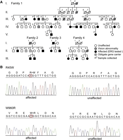
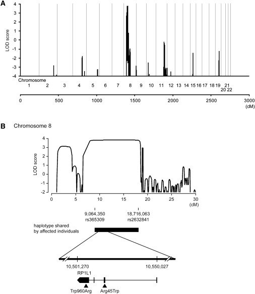
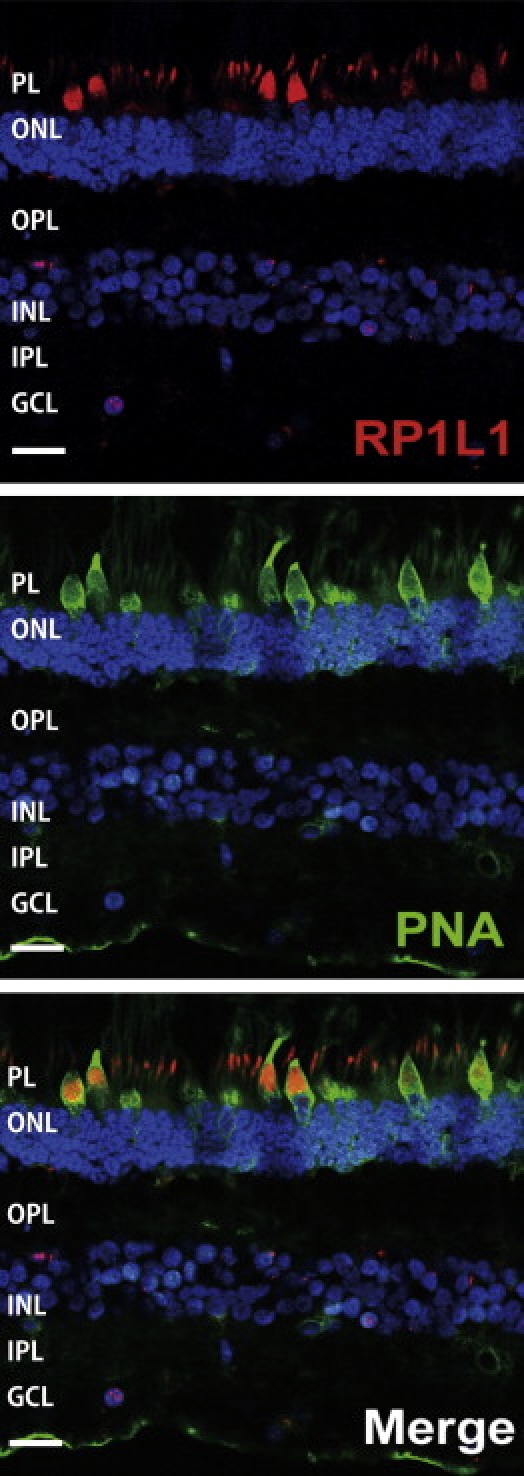
Occult macular dystrophy (OMD) is an inherited macular dystrophy characterized by progressive loss of macular function but normal ophthalmoscopic appearance. Typical OMD is characterized by a central cone dysfunction leading to a loss of vision despite normal ophthalmoscopic appearance, normal fluorescein angiography, and normal full-field electroretinogram (ERGs), but the amplitudes of the focal macular ERGs and multifocal ERGs are significantly reduced at the central retina. Linkage analysis of two OMD families was performed by the SNP High Throughput Linkage analysis system (SNP HiTLink), localizing the disease locus to chromosome 8p22-p23. Among the 128 genes in the linkage region, 22 genes were expressed in the retina, and four candidate genes were selected. No mutations were found in the first three candidate genes, methionine sulfoxide reductase A (MSRA), GATA binding 4 (GATA4), and pericentriolar material 1 (PCM1). However, amino acid substitution of p.Arg45Trp in retinitis pigmentosa 1-like 1 (RP1L1) was found in three OMD families and p.Trp960Arg in a remaining OMD family. These two mutations were detected in all affected individuals but in none of the 876 controls. Immunohistochemistry of RP1L1 in the retina section of cynomolgus monkey revealed expression in the rod and cone photoreceptor, supporting a role of RP1L1 in the photoreceptors that, when disrupted by mutation, leads to OMD. Identification of RP1L1 mutations as causative for OMD has potentially broader implications for understanding the differential cone photoreceptor functions in the fovea and the peripheral retina.
2010 The American Society of Human Genetics. Published by Elsevier Inc. All rights reserved.
Figures
References
-
- Miyake Y., Ichikawa K., Shiose Y., Kawase Y. Hereditary macular dystrophy without visible fundus abnormality. Am. J. Ophthalmol. 1989;108:292–299. - PubMed
-
- Miyake Y., Horiguchi M., Tomita N., Kondo M., Tanikawa A., Takahashi H., Suzuki S., Terasaki H. Occult macular dystrophy. Am. J. Ophthalmol. 1996;122:644–653. - PubMed
-
- Wildberger H., Niemeyer G., Junghardt A. Multifocal electroretinogram (mfERG) in a family with occult macular dystrophy (OMD) Klin. Monatsbl. Augenheilkd. 2003;220:111–115. - PubMed
-
- Piao C.H., Kondo M., Tanikawa A., Terasaki H., Miyake Y. Multifocal electroretinogram in occult macular dystrophy. Invest. Ophthalmol. Vis. Sci. 2000;41:513–517. - PubMed
-
- Brockhurst R.J., Sandberg M.A. Optical coherence tomography findings in occult macular dystrophy. Am. J. Ophthalmol. 2007;143:516–518. - PubMed
Publication types
MeSH terms
Substances
LinkOut - more resources
Full Text Sources
Medical
Molecular Biology Databases
Research Materials
