. 2010 Sep 10;87(3):436-44.
doi: 10.1016/j.ajhg.2010.08.008.

Protein tyrosine phosphatase PTPN14 is a regulator of lymphatic function and choanal development in humans

Affiliations

Protein tyrosine phosphatase PTPN14 is a regulator of lymphatic function and choanal development in humans

Audrey C Au et al. Am J Hum Genet. .

Abstract

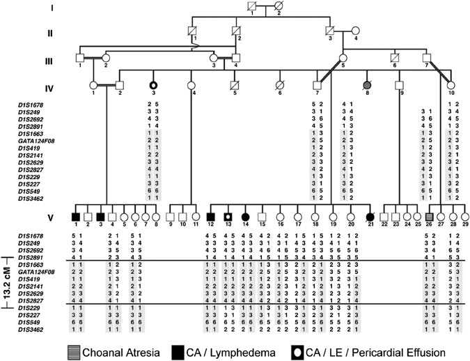
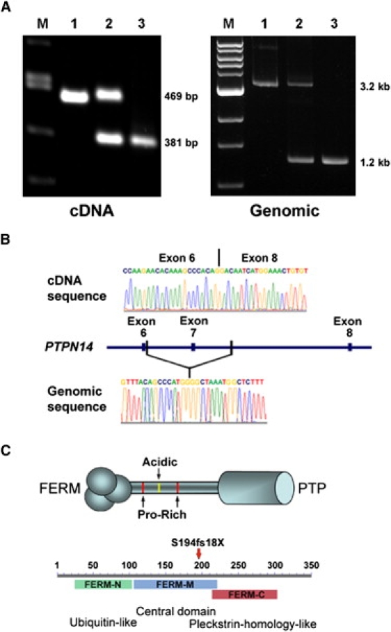
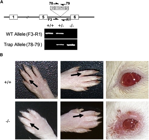
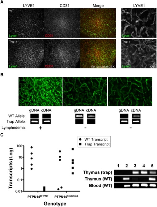
The lymphatic vasculature is essential for the recirculation of extracellular fluid, fat absorption, and immune function and as a route of tumor metastasis. The dissection of molecular mechanisms underlying lymphangiogenesis has been accelerated by the identification of tissue-specific lymphatic endothelial markers and the study of congenital lymphedema syndromes. We report the results of genetic analyses of a kindred inheriting a unique autosomal-recessive lymphedema-choanal atresia syndrome. These studies establish linkage of the trait to chromosome 1q32-q41 and identify a loss-of-function mutation in PTPN14, which encodes a nonreceptor tyrosine phosphatase. The causal role of PTPN14 deficiency was confirmed by the generation of a murine Ptpn14 gene trap model that manifested lymphatic hyperplasia with lymphedema. Biochemical studies revealed a potential interaction between PTPN14 and the vascular endothelial growth factor receptor 3 (VEGFR3), a receptor tyrosine kinase essential for lymphangiogenesis. These results suggest a unique and conserved role for PTPN14 in the regulation of lymphatic development in mammals and a nonconserved role in choanal development in humans.


Figures

Figure 1
Figure 1
Haplotype Analysis of Choanal Atresia-Lymphedema Pedigree A consanguineous pedigree is shown in which affected individuals manifest choanal atresia, lymphedema, and pericardial effusion. Individual V-26 was an infant at the time of evaluation and may have been too young to manifest lymphedema. Haplotype analysis of the critical region for choanal atresia-lymphedema syndrome on chromosome 1q32-q41 is shown. Markers inherited in homozygosity in affected individuals are boxed in gray. The telomeric boundary of the critical region is at marker D1S2891 and the centromeric boundary is at marker D1S229.
Figure 2
Figure 2
cDNA and Genomic Mutation Analysis of PTPN14 (A) Amplification products of PTPN14 cDNA (left) or genomic DNA (right) are shown. Lymphoblastoid cell total RNA was used to generate cDNA (Superscript II reverse transcriptase; Roche) as template for generating RT-PCR products spanning PTPN14 exons 3–8 from WT (left lane 1), obligate heterozygous (left lane 2), and homozygous affected individuals (left lane 3). The expected sizes of the full-length and truncated amplicon lacking exon 7 are shown at right. A 3.2 kb amplification product from genomic DNA (right lane 1) contained a ∼2 kb deletion evident in amplicons derived from heterozygous (right lane 2) and homozygous (right lane 3) individual DNA samples. (B) The absence of exon 7 was confirmed in mutant cDNA transcripts (top), and the boundaries of the genomic deletion (bottom) were defined by sequence analysis. (C) Structural features of PTPN14 include an N-terminal FERM domain, a central poorly conserved linker region with proline-rich SH3-like motifs and an acidic polyglutamate domain, and a C-terminal phosphatase domain (top). The location of the frameshift introduced by loss of exon 7 within the tripartite FERM domain is indicated by the red arrow (bottom).
Figure 3
Figure 3
A Mouse Gene Trap Model of Ptpn14 Deficiency (A) Diagram of the insertion of a gene trap viral vector into the genomic interval between exons 5 and 6 of Ptpn14. Primer pairs F3-R1 and 78-79 were used to screen for the WT and trap alleles, respectively. The results of PCR assays using tail-cut genomic DNA from WT (+/+), heterozygous (−/+), or homozygous (−/−) trap allele mice are shown below. (B) Lymphedema in the upper and lower extremities and periorbital region in adult mice homozygous for the Ptpn14 trap allele. Visible swelling of the dorsal forelimbs and/or hindlimbs and/or periorbital area was evident in a subset of animals. The distal extremities and periorbital region of a WT animal are shown (top) for comparison.
Figure 4
Figure 4
Lymphatic Hyperplasia in Symptomatic Ptpn14 Mutant Mice (A) Immunohistochemical staining of ear skin sections from WT (top) and mutant (bottom) mice. After treatment with Nair for hair removal, ear leaflets were separated and the central cartilage removed. The epidermal layer was removed by treatment with 0.5 M ammonium thiocyanate, and dermal skin was fixed with 100% acetone and 80% methanol before antibody incubations. Lymphatic vessels were stained with rabbit anti-LYVE1 (Upstate) followed by anti-rabbit Alex Fluor 488 (Molecular Probes), and blood vessels were visualized with panendothelial rat anti-CD31 (BD Biosciences) followed by anti-rat Alexa Fluor 594 (Molecular Probes). Images were captured with a Zeiss Axiophot2 fluorescence microscope after mounting (Dako Cytomation). Immunostaining of the lymphatic endothelial marker LYVE1 (green) and the vascular endothelial predominant marker CD31 (red) are shown at low magnification (25×). A higher-magnification view (100×) of LYVE1 staining is shown at right. The sections from mutant animals have an increased density of LYVE1-positive lymphatic capillaries compared to WT ear sections. (B) Ear sections from symptomatic mutant (left), WT (center), and nonsymptomatic mutant (right) mice are shown with confirmatory genotyping (gDNA) and expression analysis (cDNA) for Ptpn14 WT and trap alleles shown below. Genomic DNA was obtained from tail cuts, and cDNA was obtained from peripheral blood. The Ptpn14 trap transcript was amplified with primers specific for exon 5 and the β-geo open reading frame. WT transcripts arising by skipping of the trap exon were detected with the use of primers flanking the exon 5–6 splice junction. WT transcripts were readily detected with cDNA derived from asymptomatic mice but not with that from symptomatic mice. (C) Real-time quantitative PCR with thymocyte-derived cDNA from WT and asymptomatic trapped animals for the calculation of generated WT and trapped transcripts. 20 ng of cDNA and 0.2 μM of each primer (Table S1) with SYBR Green Master (Rox) (Roche) were amplified in an ABI PRISM 7900HT detection system for 40 cycles in triplicate. Transcript abundance was calculated with the formula 2500 × 1.93̂((mean Gapdh Ct)−(mean transcript Ct)), with Ct as the threshold cycle. The right panel shows differential splicing of WT transcripts in asymptomatic trapped animals in thymocyte- versus leukocyte-derived cDNA. WT and trap products from qPCR in the left panel were analyzed by gel electrophoresis. Lanes: 1, negative control; 2, WT; 3–5, homozygous Ptpn14trap. Trap amplicons derived from the thymus were present in all mutant samples but not in the WT sample (top panel). WT amplicons were present in only one (lane 5) of the three mutant samples derived from the thymus (middle panel) but in all samples derived from blood (bottom panel).
Figure 5
Figure 5
PTPN14 Can Modulate VEGFR3 Receptor Activation in a Specific Fashion (A) cDNA and total protein lysates were prepared from human foreskin lymphatic endothelial cells (LECs) for RT-PCR and immunoblot analyses. PTPN14 polyclonal rabbit primary antisera (a generous gift from Y. Khew-Goodall) were raised against a peptide corresponding to PTPN14 residues 683–696 as described previously, and subsequent aliquots were generated against this same epitope in rabbits. PTPN14 transcription (top) and protein expression (bottom) were confirmed in LECs, absent in HEK293 cells (−), and present in HEK293 cells expressing Flag-tagged PTPN14 (+). (B) HEK293 cells were cotransfected with combinations of V5-tagged VEGFR3, V5-tagged EB1, and FLAG-tagged PTPN14 as indicated at right. Confluent HEK293 cell monolayers were washed with PBS on ice and then scraped with lysis buffer (20 mM Tris pH 7.6, 150 mM NaCl, 50 mM NaF, 1 mM Na3VO4, 1 mM EDTA, 1% [v/v] Triton X-100, 0.5% [w/v] sodium deoxycholate, 0.1% [w/v] SDS) and protease inhibitors (Complete Protease Inhibitor Tablets, Roche). Lysates were immunoprecipitated with mouse anti-V5 antibody (Invitrogen), and coimmunoprecipitation of FLAG-tagged PTPN14 (top row) was detected by immunoblotting with anti-PTPN14 antibody. Blots were stripped and reprobed with anti-V5 antibody to confirm the immunoprecipitation of V5-tagged VEGFR3 (second row) and V5-tagged EB1 (third row). PTPN14 coimmunoprecipitated with V5-tagged VEGFR3, but not with a control protein, V5-tagged EB1, under basal conditions in serum-replete medium (lanes 3 and 4). Human recombinant vascular endothelial growth factor-C (VEGFC; R&D Systems) stimulation of transfected cells resulted in a more robust interaction relative to serum-starved cells (lanes 5 and 6). Experiments were repeated three to five times, and representative data are shown.

References

    1. Cueni L.N., Detmar M. The lymphatic system in health and disease. Lymphat. Res. Biol. 2008;6:109–122. - PMC - PubMed
    1. Karpanen T., Alitalo K. Molecular biology and pathology of lymphangiogenesis. Annu. Rev. Pathol. 2008;3:367–397. - PubMed
    1. Tammela T., Alitalo K. Lymphangiogenesis: Molecular mechanisms and future promise. Cell. 2010;140:460–476. - PubMed
    1. Ferrell R.E., Levinson K.L., Esman J.H., Kimak M.A., Lawrence E.C., Barmada M.M., Finegold D.N. Hereditary lymphedema: evidence for linkage and genetic heterogeneity. Hum. Mol. Genet. 1998;7:2073–2078. - PubMed
    1. Irrthum A., Karkkainen M.J., Devriendt K., Alitalo K., Vikkula M. Congenital hereditary lymphedema caused by a mutation that inactivates VEGFR3 tyrosine kinase. Am. J. Hum. Genet. 2000;67:295–301. - PMC - PubMed

MeSH terms

Substances

Associated data