

Protective role of interleukin-10 in ozone-induced pulmonary inflammation
- PMID: 20826374
- PMCID: PMC3002191
- DOI: 10.1289/ehp.1002182
Protective role of interleukin-10 in ozone-induced pulmonary inflammation
Abstract
Background: The mechanisms underlying ozone (O₃)-induced pulmonary inflammation remain unclear. Interleukin-10 (IL-10) is an anti-inflammatory cytokine that is known to inhibit inflammatory mediators.
Objectives: We investigated the molecular mechanisms underlying interleuken-10 (IL-10)-mediated attenuation of O₃-induced pulmonary inflammation in mice.
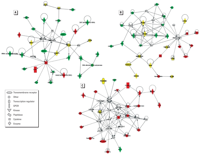
Methods: Il10-deficient (Il10(-/-)) and wild-type (Il10(+/+)) mice were exposed to 0.3 ppm O₃ or filtered air for 24, 48, or 72 hr. Immediately after exposure, differential cell counts and total protein (a marker of lung permeability) were assessed from bronchoalveolar lavage fluid (BALF). mRNA and protein levels of cellular mediators were determined from lung homogenates. We also used global mRNA expression analyses of lung tissue with Ingenuity Pathway Analysis to identify patterns of gene expression through which IL-10 modifies O₃-induced inflammation.
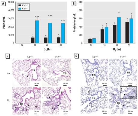
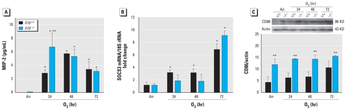
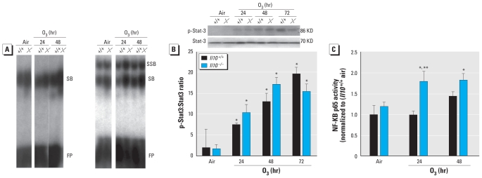
Results: Mean numbers of BALF polymorphonuclear leukocytes (PMNs) were significantly greater in Il10(-/-) mice than in Il10(+/+) mice after exposure to O₃ at all time points tested. O₃-enhanced nuclear NF-κB translocation was elevated in the lungs of Il10(-/-) compared with Il10(+/+) mice. Gene expression analyses revealed several IL-10-dependent and O₃-dependent mediators, including macrophage inflammatory protein 2, cathepsin E, and serum amyloid A3.
Conclusions: Results indicate that IL-10 protects against O₃-induced pulmonary neutrophilic inflammation and cell proliferation. Moreover, gene expression analyses identified three response pathways and several genetic targets through which IL-10 may modulate the innate and adaptive immune response. These novel mechanisms of protection against the pathogenesis of O₃-induced pulmonary inflammation may also provide potential therapeutic targets to protect susceptible individuals.
Figures
References
-
- Akdis CA, Joss A, Akdis M, Blaser K. Mechanism of IL-10-induced T cell inactivation in allergic inflammation and normal response to allergens. Int Arch Allergy Immunol. 2001;124((1–3)):180–182. - PubMed
-
- American Physiological Society. Appendix C. Guiding Principles for Research Involving Animals and Human Beings. Recommendations From The Revised Declaration of Helsinki by the World Medical Association Regarding Human Subjects. 2002. [(accessed 21 September 2010)]. Available: http://www.the-aps.org/about/opguide/appendix.htm#guiding.
-
- Berlato C, Cassatella MA, Kinjyo I, Gatto L, Yoshimura A, Bazzoni F. Involvement of suppressor of cytokine signaling-3 as a mediator of the inhibitory effects of IL-10 on lipopolysaccharide-induced macrophage activation. J Immunol. 2002;168((12)):6404–6411. - PubMed
-
- Bhalla DK, Reinhart PG, Bai C, Gupta SK. Amelioration of ozone-induced lung injury by antitumor necrosis factor-alpha. Toxicol Sci. 2002;69((2)):400–408. - PubMed
Publication types
MeSH terms
Substances
Grants and funding
LinkOut - more resources
Full Text Sources
Medical
Molecular Biology Databases