

Rho-associated kinase-dependent contraction of stress fibres and the organization of focal adhesions
- PMID: 20826475
- PMCID: PMC3030825
- DOI: 10.1098/rsif.2010.0419
Rho-associated kinase-dependent contraction of stress fibres and the organization of focal adhesions
Abstract
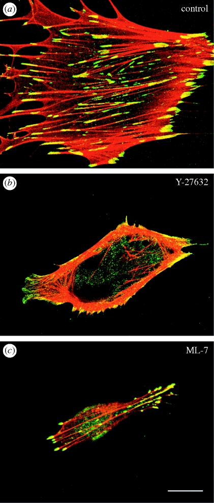
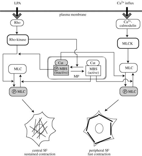
Stress fibres and associated focal adhesions in cells constitute a contractile apparatus that regulates cell motility and contraction. Rho-kinase, an effector molecule of small GTPases, regulates non-muscle cell motility and contractility. Rho-kinase mediates the contraction of stress fibres in a Ca(2+)-independent manner, and is responsible for slower and more finely tuned contraction of stress fibres than that regulated by myosin light chain kinase activity in living cells. The specific inhibition of the Rho-kinase activity causes cells to not only lose their stress fibres and focal adhesions, but also to appear to lose their cytoplasmic tension. Activated Rho-kinase is also involved in the organization of newly formed stress fibres and focal adhesions in living cells.
Figures
References
-
- Byers H. R., Fujiwara K. 1982. Stress fibers in cells in situ: immunofluorescence visualization with antiactin, antimyosin, and anti-alpha-actinin. J. Cell Biol. 93, 804–811 10.1083/jcb.93.3.804 (doi:10.1083/jcb.93.3.804) - DOI - PMC - PubMed
-
- White G. E., Gimbrone M. A. J., Fujiwara K. 1983. Factors influencing the expression of stress fibers in vascular endothelial cells in situ. J. Cell Biol. 97, 416–424 10.1083/jcb.97.2.416 (doi:10.1083/jcb.97.2.416) - DOI - PMC - PubMed
-
- Sugimoto K., Fujii S., Yamashita K. 1991. Expression of stress fibers in bullfrog mesothelial cells in response to tension. Exp. Cell Res. 196, 353–361 10.1016/0014-4827(91)90271-U (doi:10.1016/0014-4827(91)90271-U) - DOI - PubMed
-
- Murakami T., Ishikawa H. 1991. Stress fibers in situ in proximal tubules of the rat kidney. Cell Struct. Funct. 16, 231–240 10.1247/csf.16.231 (doi:10.1247/csf.16.231) - DOI - PubMed
Publication types
MeSH terms
Substances
LinkOut - more resources
Full Text Sources
Miscellaneous