Review
. 2010 Nov;2(11):a000547.
doi: 10.1101/cshperspect.a000547. Epub 2010 Sep 8.

Nuclear lamins

Affiliations
Review

Nuclear lamins

Thomas Dechat et al. Cold Spring Harb Perspect Biol. 2010 Nov.

Abstract

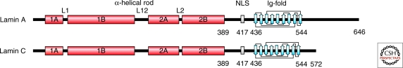
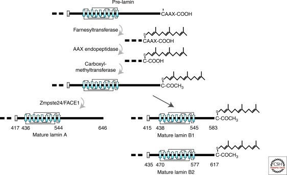
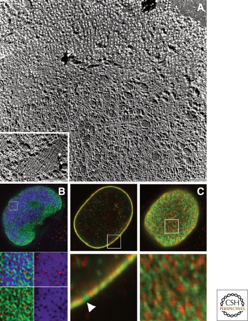
The nuclear lamins are type V intermediate filament proteins that are critically important for the structural properties of the nucleus. In addition, they are involved in the regulation of numerous nuclear processes, including DNA replication, transcription and chromatin organization. The developmentally regulated expression of lamins suggests that they are involved in cellular differentiation. Their assembly dynamic properties throughout the cell cycle, particularly in mitosis, are influenced by posttranslational modifications. Lamins may regulate nuclear functions by direct interactions with chromatin and determining the spatial organization of chromosomes within the nuclear space. They may also regulate chromatin functions by interacting with factors that epigenetically modify the chromatin or directly regulate replication or transcription.


Figures

Figure 1.
Figure 1.
Structure of nuclear lamins. Schematic drawing of mature lamin A and lamin C polypeptide chains. The lamin structure consists of a short amino terminal head domain, a central α-helical rod domain (red), and the carboxy-terminal domain containing the NLS and the Ig-fold (blue; the nine β-strands of the Ig-fold motif are depicted). Modified, with permission, from (Dechat et al. 2008b).
Figure 2.
Figure 2.
Assembly of the nuclear lamins in vitro. Lamins self assemble to form dimers (A) which then join to form linear head-to-tail polymers (protofilaments) (B). Bar = 100 nm; electron micrograph of rotary shadowed chicken lamin B2. These protofilaments further assemble into “beaded” filaments or fibers (C) which in turn associate laterally into thicker fibers (D), and eventually into paracrystalline arrays (E); C,D are negatively stained electron microscope preparations. Reprinted from Stuurman et al. (1998) with permission from Elsevier.
Figure 3.
Figure 3.
Posttranslational processing of the carboxyl terminus of prelamins A, B1, and B2. Processing takes place in a series of steps: (1) addition of a farnesyl group to the cysteine residue of the –CAAX box of pre-lamin A, prelamin B1 and prelamin B2 by a farnesyltransferase; (2) removal of the last three residues (−AAX) by an AAX endopeptidase; (3) methylation of the terminal carboxylic acid group (−COOH) by a carboxyl methyltransferase; (4) removal of the carboxyl terminal 15 amino acids of lamin A with the farnesyl attached by the metalloprotease Zmpste24/FACE1. This last proteolysis step does not occur on B-type lamins and therefore they remain farnesylated. Modified, with permission, from Dechat et al. (2008b).
Figure 4.
Figure 4.
The nuclear lamins form a meshwork of filaments within the lamina. (A) Spread nuclear envelope from Xenopus oocytes after detergent extraction and preparation for transmission electron microscopy by freeze-drying/unidirectional metal shadowing. The micrograph shows the nuclear lamina meshwork partially studded with nuclear pore complexes. (Inset) Higher-magnification view of a particularly well-preserved area clearly shows the near-tetragonal lamina meshwork. Bars, 1 µm. Reprinted from Stuurman et al. (1998), with permission from Elsevier. (B) Structured illumination microsopy (SIM) reveals that there is an irregular meshwork of nuclear lamin B as revealed by immunofluorescence (green). This preparation is also stained with antibodies directed against nuclear pores and is stained with DAPI for DNA/chromatin. Pores (red), DAPI (blue). From Schermelleh et al. (2008). Reprinted with permission from AAAS. (C) Confocal immunofluorescence localization of lamin A/C (green) and lamin B1 (red) in HeLa cells. Lamins A/C and B1 in a single nucleus are seen in an equatorial section (left panels) and the nuclear surface (right panels). The areas indicated by white squares in the top panels are enlarged fivefold in the lower panels. These images demonstrate that lamins form mainly separate networks with some overlapping regions. Bar, 5 µm. Adapted, with permission, from Shimi et al. (2008).
Figure 5.
Figure 5.
Association of lamin A with chromosomes during mitosis. HeLa cells expressing GFP-lamin A were followed by time-lapse microscopy from the metaphase/anaphase transition (far left panels) into early G1 (far right panels). GFP-lamin A first associates with the core regions of chromosomes during telophase and spreads to cover the entire chromatin surface by early G1. DIC images of the same series are shown in the bottom row (Dechat et al. 2007).
Figure 6.
Figure 6.
The nuclear lamina is a fibrous network (fibrous lamina) located between peripheral heterochromatin and the inner nuclear membrane. A transmission electron micrograph of a thin section showing a portion of a smooth muscle nucleus in guinea pig epedidymis. Adapted from Fawcett (1966), copyright 1995, Wiley-Liss, Inc. Reprinted with permission of John Wiley & Sons, Inc. American Journal of Anatomy, Vol. 119, No. 1, pg. 140.
Figure 7.
Figure 7.
The association of the nuclear lamina with chromatin in normal and progeria patients' cells. (A) Thin section electron micrograph of a late passage blebbed nucleus in an HGPS patient's skin fibroblast (left and center panels) and normal human foreskin fibroblasts (right panel). A high-magnification view of the nuclear envelope in a normal human foreskin fibroblast shows a normal array of heterochromatin adjacent to the nuclear envelope, making any lamina structure difficult to detect (right panel). A higher-magnification view of a HGPS cell showing a loss of peripheral heterochromatin and a prominent electron-dense lamina region associated with the inner nuclear envelope membrane (the nucleus is to the left in center and right panels) (Goldman et al. 2004). (B) Alterations of histone methylation patterns in HGPS fibroblasts. Normal and HGPS fibroblasts from female donors were double-labeled with antibodies against lamins A/C (red) and trimethylated Lys 9 in histone H3 (H3K9me3), Lys 27 in histone H3 (H3K27me3), or Lys 20 in histone H4 (H4K20me3) (all green). Note the decrease of H3K9me3 and H3K27me3 and the increase of H4K20me3 in the lobulated HGPS nuclei compared with normal nuclei. The decrease in H3K27me3 is best observed at the inactive X chromosome, which is normally enriched in this histone modification (see arrowhead in center left panel). Bars, 10 µm. Reprinted, with permission, from Dechat et al. (2008b).
Figure 8.
Figure 8.
Localization of specific gene rich chromosomal regions in nuclear blebs in LB1-silenced HeLa cells. Chromosomes are detected by fluorescence in situ hybridization. DNA is counterstained with Hoechst 33258 (blue). Chromosome 6p (or 19), and chromosome 6q (or 18) are shown in green and red, respectively. Bars, 5 µm. Adapted, with permission, from Shimi et al. (2008).
Figure 9.
Figure 9.
Mislocalization of heterochromatin and centromeres in HGPS E145K cells. Control and E145K patient cells were stained with anti-lamin A (red), CREST antiserum (green), and Hoechst (white). Maximum projections of series of z-sections spanning the entire nucleus and side projections are shown. Centromeres are clustered in the central region of the nucleus in E145K cells, while they are either closely associated with the peripheral lamina region or elsewhere in the nucleoplasm in control cells. Inset shows an example of the close association of one centromere with the lamina in a single confocal section. In the right hand panels, side projections from merged images are shown. Centromeres are distributed throughout the nuclei in normal cells. In E145K cells centromeres are clustered in the middle of one region of the nucleus (Taimen et al. 2009a).

References

    1. Aaronson RP, Blobel G 1975. Isolation of nuclear pore complexes in association with a lamina. Proc Natl Acad Sci 72: 1007–1011 - PMC - PubMed
    1. Adolph KW 1987. ADPribosylation of nuclear proteins labeled with [3H]adenosine: changes during the HeLa cycle. Biochim Biophys Acta 909: 222–230 - PubMed
    1. Aebi U, Cohn J, Buhle L, Gerace L 1986. The nuclear lamina is a meshwork of intermediate-type filaments. Nature 323: 560–564 - PubMed
    1. Akter R, Rivas D, Geneau G, Drissi H, Duque G 2009. Effect of lamin A/C knockdown on osteoblast differentiation and function. J Bone Miner Res 24: 283–293 - PubMed
    1. Andres V, Gonzalez JM 2009. Role of A-type lamins in signaling, transcription, and chromatin organization. J Cell Biol 187: 945–957 - PMC - PubMed

Publication types

LinkOut - more resources