. 2010 Sep 8;30(36):11926-37.
doi: 10.1523/JNEUROSCI.5642-09.2010.

The role of contralesional dorsal premotor cortex after stroke as studied with concurrent TMS-fMRI

Affiliations

The role of contralesional dorsal premotor cortex after stroke as studied with concurrent TMS-fMRI

Sven Bestmann et al. J Neurosci. .

Abstract

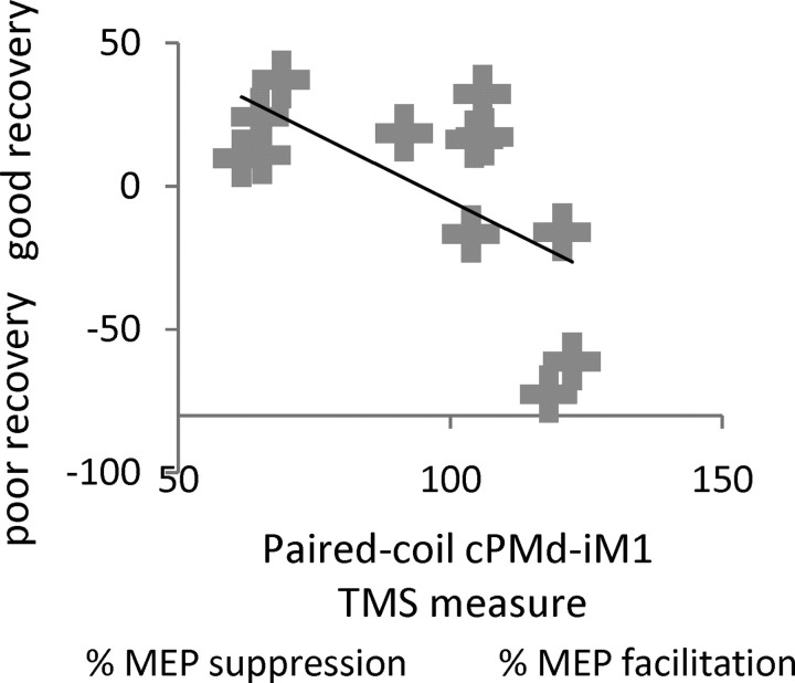
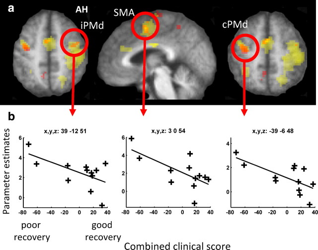
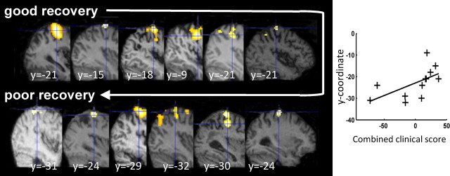
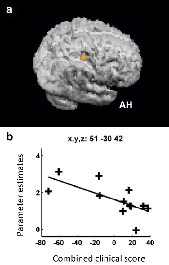
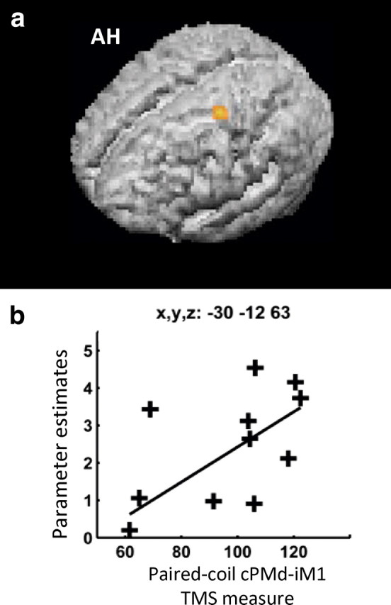
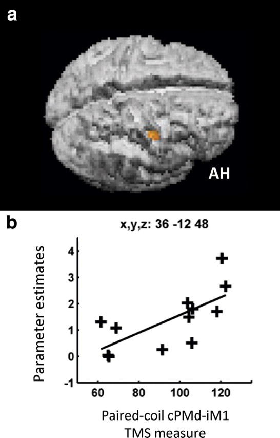
Contralesional dorsal premotor cortex (cPMd) may support residual motor function following stroke. We performed two complementary experiments to explore how cPMd might perform this role in a group of chronic human stroke patients. First, we used paired-coil transcranial magnetic stimulation (TMS) to establish the physiological influence of cPMd on ipsilesional primary motor cortex (iM1) at rest. We found that this influence became less inhibitory/more facilitatory in patients with greater clinical impairment. Second, we applied TMS over cPMd during functional magnetic resonance imaging (fMRI) in these patients to examine the causal influence of cPMd TMS on the whole network of surviving cortical motor areas in either hemisphere and whether these influences changed during affected hand movement. We confirmed that hand grip-related activation in cPMd was greater in more impaired patients. Furthermore, the peak ipsilesional sensorimotor cortex activity shifted posteriorly in more impaired patients. Critical new findings were that concurrent TMS-fMRI results correlated with the level of both clinical impairment and neurophysiological impairment (i.e., less inhibitory/more facilitatory cPMd-iM1 measure at rest as assessed with paired-coil TMS). Specifically, greater clinical and neurophysiological impairment was associated with a stronger facilitatory influence of cPMd TMS on blood oxygenation level-dependent signal in posterior parts of ipsilesional sensorimotor cortex during hand grip, corresponding to the posteriorly shifted sensorimotor activity seen in more impaired patients. cPMd TMS was not found to influence activity in other brain regions in either hemisphere. This state-dependent influence on ipsilesional sensorimotor regions may provide a mechanism by which cPMd supports recovered function after stroke.


Figures

Figure 1.
Figure 1.
Experimental setup and main effects of the grip task. a, Left, Photograph of grip-force manipulandum. Right, Screens displayed during scanning for a grip trial (arrow shown at bottom of thermometer-like visual display) and a no-grip trial (central cross being presented instead of the arrow). During grip trials, a yellow target bar indicated the required force level, as shown in the schematic example. The actual force exerted by the affected hand was indicated by red shading of the thermometer-like display and the white arrow pointed to the paretic hand to indicate that active grip was required. Participants were instructed to generate a nonballistic force matching the displayed target bar, using a gentle pace without major corrective movements (see main text). In all trials, TMS (5 pulses, 11 Hz) was applied unpredictably to contralesional PMd at one of two intensities (110% resting motor threshold or 70% active motor threshold) 900 ms after presentation of the force target level (or cross) visual instruction. b, Hand grip-related activity, regardless of TMS. The results of the group random-effects analysis are projected onto the rendered averaged structural scans from all patients. The height threshold was set at t > 3.5, uncorrected for multiple comparisons across whole brain, and the extent (or cluster) threshold set at p < 0.05, corrected for multiple comparisons across the whole brain. AH, Affected hemisphere.
Figure 2.
Figure 2.
Scatterplot showing the correlation, with regression line, between the combined clinical score and the interhemispheric cPMd-iM1 influence measured with paired-coil TMS (conditioned MEP/unconditioned MEP as a percentage) in each patient. For the combined clinical score (along the y-axis), a higher value indicates better residual motor function. This measure correlated with the value of the interhemispheric cPMd-iM1 influence shown along the x-axis; a better motor recovery was associated with a physiological inhibitory effect, whereas poorer recovery was associated with less interhemispheric inhibition or even facilitation (i.e., paired-coil effects of >100%, as for the rightmost cases).
Figure 3.
Figure 3.
Hand grip-related fMRI activity correlations with residual motor function. a, SPM for the main effect of hand grip (minus rest) is shown in yellow, overlaid onto the mean normalized T1-weighted structural image from all participants. Activation clusters in which a significant relationship between hand grip-related activity and the combined clinical score was observed are shown in orange (p < 0.05, corrected, for multiple comparisons across the brain). b, Parameter estimates from each individual patient for the main effect of hand grip (minus rest; shown along the y-axis) plotted against the combined clinical score from each patient (along the x-axis) for the circled regions in a, at the coordinates listed. iPMd, Ipsilesional PMd; SMA, supplementary motor area; AH, affected hemisphere.
Figure 4.
Figure 4.
Relationship between the ipsilesional peak hand grip-related signal change and the combined clinical score. The SPM for the main effect of hand grip is overlaid on the individual structural scan of each patient. A posterior shift of activity in ipsilesional cortex was observed, with less well recovered patients exhibiting progressively more posterior peak activity. The y-coordinate from each individual patient for the peak activity for the main effect of grip versus rest (shown along the y-axis) is plotted against the combined clinical score from each patient (along the x-axis).
Figure 5.
Figure 5.
a, The facilitatory influence of contralesional PMd during hand grip (as measured with concurrent TMS-fMRI) correlated with combined clinical score in this ipsilesional cluster extending across posterior sensorimotor cortical regions. b, SPM for the interaction term TMShigh (grip–rest) > TMSlow (grip–rest) overlaid on the rendered mean structural scan from all patients. The influence of cPMd on this cluster [assessed by the parameter estimates for TMShigh (grip–rest) > TMSlow (grip–rest)] are plotted against the combined clinical score for each patient. AH, Affected hemisphere.
Figure 6.
Figure 6.
a, cPMd-iM1 interhemispheric influences at rest (as revealed by paired-coil TMS) correlated with hand grip-related activity in contralesional premotor cortex. Hand grip-related activity at the identified stimulation site in cPMd correlated with the separately measured interhemispheric paired-coil cPMd-iM1 influence (p < 0.05, corrected, for multiple comparisons across the brain). The SPM for this effect is projected onto the rendered, average, normalized, T1-weighted structural image from all participants. b, Parameter estimates for the main effect of hand grip minus rest (shown along the y-axis) are plotted in a patient-by-patient manner against the interhemispheric cPMd-iM1 influence (along the x-axis) are shown in the inlay. AH, Affected hemisphere.
Figure 7.
Figure 7.
Brain regions in which the influence of cPMd during hand grip (as measured with concurrent TMS-fMRI) was greater when cPMd had a less inhibitory/more facilitatory affect on ipsilesional M1 (as measured with paired-coil TMS). a, The SPM for the correlation seen in ipsilesional posterior central sulcus, BA4p, is overlaid on the rendered mean structural scan from all patients. b, Each patient's parameter estimate for the interaction term TMShigh (grip–rest) > TMSlow (grip–rest) in ipsilesional posterior central sulcus, BA4p, plotted against the paired-coil measure of the interhemispheric cPMd-iM1 influence. AH, Affected hemisphere.

References

    1. Amassian VE, Stewart M. Motor cortical and other cortical interneuronal networks that generate very high frequency waves. Suppl Clin Neurophysiol. 2003;56:119–142. - PubMed
    1. Amiez C, Kostopoulos P, Champod AS, Petrides M. Local morphology predicts functional organization of the dorsal premotor region in the human brain. J Neurosci. 2006;26:2724–2731. - PMC - PubMed
    1. Andersson JL, Hutton C, Ashburner J, Turner R, Friston K. Modeling geometric deformations in EPI time series. Neuroimage. 2001;13:903–919. - PubMed
    1. Baudewig J, Paulus W, Frahm J. Artifacts caused by transcranial magnetic stimulation coils and EEG electrodes in T(2)*-weighted echo-planar imaging. Magn Reson Imaging. 2000;18:479–484. - PubMed
    1. Baudewig J, Siebner HR, Bestmann S, Tergau F, Tings T, Paulus W, Frahm J. Functional MRI of cortical activations induced by transcranial magnetic stimulation (TMS) Neuroreport. 2001;12:3543–3548. - PubMed

Publication types

MeSH terms