. 2010 Sep 8;30(36):12005-19.
doi: 10.1523/JNEUROSCI.2208-10.2010.

Reduced survival of motor neuron (SMN) protein in motor neuronal progenitors functions cell autonomously to cause spinal muscular atrophy in model mice expressing the human centromeric (SMN2) gene

Affiliations

Reduced survival of motor neuron (SMN) protein in motor neuronal progenitors functions cell autonomously to cause spinal muscular atrophy in model mice expressing the human centromeric (SMN2) gene

Gyu-Hwan Park et al. J Neurosci. .

Abstract

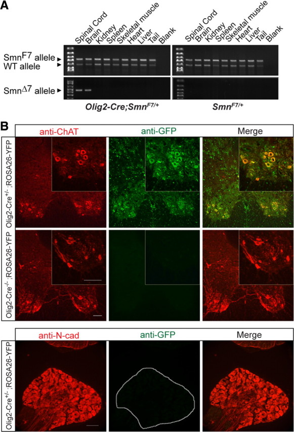
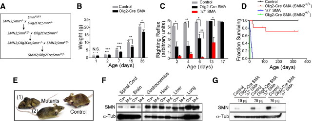
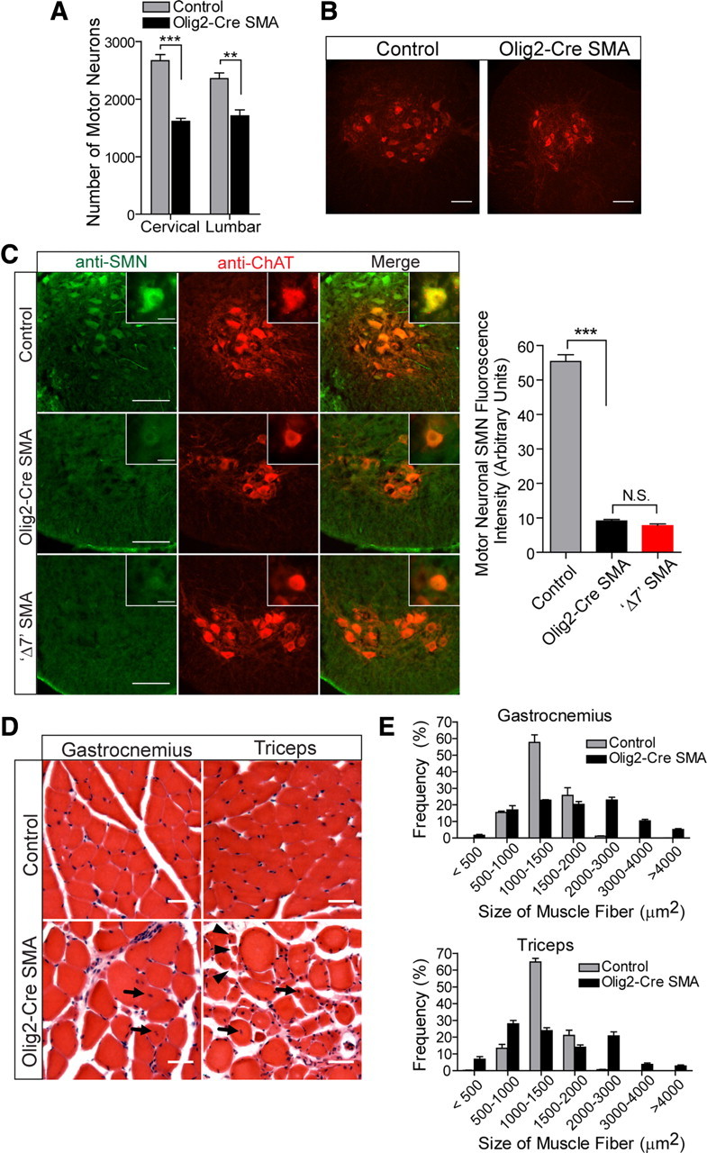
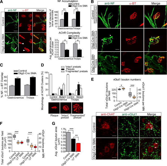
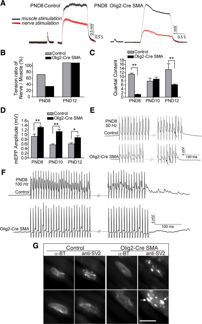
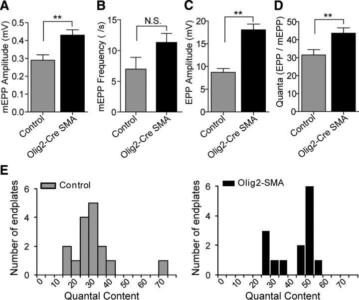
Spinal muscular atrophy (SMA) is a common (approximately 1:6400) autosomal recessive neuromuscular disorder caused by a paucity of the survival of motor neuron (SMN) protein. Although widely recognized to cause selective spinal motor neuron loss when deficient, the precise cellular site of action of the SMN protein in SMA remains unclear. In this study we sought to determine the consequences of selectively depleting SMN in the motor neurons of model mice. Depleting but not abolishing the protein in motor neuronal progenitors causes an SMA-like phenotype. Neuromuscular weakness in the model mice is accompanied by peripheral as well as central synaptic defects, electrophysiological abnormalities of the neuromuscular junctions, muscle atrophy, and motor neuron degeneration. However, the disease phenotype is more modest than that observed in mice expressing ubiquitously low levels of the SMN protein, and both symptoms as well as early electrophysiological abnormalities that are readily apparent in neonates were attenuated in an age-dependent manner. We conclude that selective knock-down of SMN in motor neurons is sufficient but may not be necessary to cause a disease phenotype and that targeting these cells will be a requirement of any effective therapeutic strategy. This realization is tempered by the relatively mild SMA phenotype in our model mice, one explanation for which is the presence of normal SMN levels in non-neuronal tissue that serves to modulate disease severity.


Figures

Figure 1.
Figure 1.
Specificity of Olig2-Cre driven SmnF7 recombination in nervous tissue. A, Primers specific for the SmnΔ7 allele detect the recombination event in genomic DNA isolated from spinal cord and brain tissue alone of adult (PND30) Olig2-Cre;SmnF7/+ but not SmnF7/+mice. The presence of bands corresponding to the SmnF7 allele in nervous tissue of the double transgenics is reflective of non-Olig2-Cre expressing cells. Amplification of the wild-type allele serves as an internal control. B, Detection of GFP fluorescence in ChAT-positive ventral horn motor neurons of Olig2-Cre expressing ROSA26-flox-STOP-flox-YFP mice is evidence of strong expression of Cre-recombinase in these cells. Absence of detectable GFP fluorescence in the neural cadherin-expressing DRG sensory neurons of the mice indicates that Olig2-Cre expression is not pan-neuronal. ROSA26-flox-STOP-flox-YFP mice lacking the Olig2-Cre transgene fail to express GFP fluorescence in ventral horn cells. Scale bars, 100 μm.
Figure 2.
Figure 2.
Olig2-Cre SMA mutants express low SMN protein in nervous tissue and exhibit a neuromuscular phenotype. A, Genetic crosses to generate Olig2-Cre SMA mutant mice. B, A weight difference becomes apparent in Olig2-Cre SMA mutants at PND2 and remains significant (*p < 0.05; **p < 0.01; ***p < 0.001) into adulthood; n ≥ 10 for each genotype. C, Early and severe neuromuscular weakness in Olig2-Cre SMA mutants is mitigated in an age-dependent manner, whereas the phenotype remains obvious as the disease progresses in Δ7 SMA mouse mutants and is considerably worse (mean difference, 3.001, p < 0.001, one-way ANOVA) than the former cohort at PND6; n ≥ 12 for each genotype. D, An increase in disease severity of Δ7 SMA mutants expressing ubiquitously low SMN as compared to Olig2-Cre SMA mice selectively depleted for protein in nervous tissue. Kaplan–Meier survival curves, censored at PND340, are significantly different between the two mutant groups (log-rank test, χ2 = 26.09, p < 0.0001, n ≥ 10 for each genotype). Additionally, Olig2-Cre SMA mutants (n = 11) hemizygous for SMN2 have a significantly reduced survival (mean survival, 1.81 ± 0.7 d) compared to Olig2-Cre mutants homozygous for the transgene. Note: *p < 0.01; **p < 0.001. E, Gross phenotype of Olig2-Cre SMA mutants displaying a relatively severe (1) and a more mild (2) form of neuromuscular disease. Severe mutants often display a prominent kyphosis (arrow) of the spine. F, Severe and selective reduction of SMN protein in nervous tissue of Olig2-Cre SMA mutants (Mut) and control (Con) littermates. α-Tub, α-Tubulin. G, Estimation of SMN in spinal cord extracts (10, 20, and 30 μg) of PND15 Δ7 SMA, Olig2-Cre SMA, and control animals indicates a significant depletion of protein in the mutants. As expected, selective depletion in motor neuronal progenitors results in greater levels in Olig2-Cre mutant spinal cords compared to Δ7 SMA tissue. Control: SMN2+/+;Olig2-Cre+/−;SmnF7/+; Olig2-Cre SMA: SMN2+/+;Olig2-Cre+/−;SmnF7/−; Δ7 SMA: SMN2+/+;SMNΔ7+/+;Smn−/−.
Figure 3.
Figure 3.
Neuromuscular pathology of Olig2-Cre SMA mutants. A, B, Motor neuron loss is significant in the lumbar (**p < 0.01, n > 5, t test) and cervical (***p < 0.001, n > 5, t test) spinal cords of mutant animals. C, Surviving (ChAT positive) motor neurons of Olig2-Cre SMA mutants express levels of SMN protein comparable to those of Δ7 SMA mice and markedly less than cells in control littermates. Insets depict enlarged examples of individual motor neurons in the different mice. Staining intensity of Olig2-Cre motor neurons was significantly reduced compared to those of control mice but no different from those of Δ7 SMA mutants (n ≥ 50 cells; ***p < 0.001 between controls and mutants; N.S.p > 0.05 between Olig2-Cre and Δ7 mutants, one-way ANOVA). D, Muscle pathology of Olig2-Cre SMA mutants, as assessed in transverse section of H&E-stained proximal and distal muscles, is characterized by atrophic fibers (arrowheads) centrally located nuclei (arrows). and hypertrophy. E, Frequency distribution of the area of individual muscle fibers indicates significant hypertrophy of both proximal and distal muscle groups in Olig2-Cre SMA mutants (n ≥ 4 of each genotype). Scale bars, B, 100 μm; C, 100 μm, insets, 25 μm, D, 50 μm.
Figure 4.
Figure 4.
Peripheral and central synaptic defects in Olig2-Cre SMA mutants. A, Selectively depleting SMN in motor neurons is sufficient to cause defects of presynaptic as well as postsynaptic specializations of the neuromuscular synapses by 2 weeks of age. Nerve terminals as seen in the triceps appear infiltrated with abnormal levels of NF protein (arrows). On average, NF fluorescence intensity was twice as great in both proximal and distal muscles of mutant animals, while AChR clusters at the postsynapse fail to develop into so-called pretzels (arrowheads). NF accumulation: **p < 0.01; N = 3 mice, n ≥ 40 NMJs; AChR complexity: *p < 0.05; N = 3 mice, n ≥ 70 NMJs, t test in both instances. B, NMJs in distal muscles (gastrocnemius) of adult Olig2-Cre SMA mice mature but are predominantly fragmented and continue to display prominent NF swellings (arrowheads) that often fail to overlap with AChRs (arrows). Conversely, NMJs of the proximal triceps appear mostly poorly developed with fewer perforations and NF aggregates. C, Quantification of NF accumulation in presynapses of adult mutant muscle based upon the degree of overlap between NF-positive nerve terminals and α-BT staining AChRs, p < 0.01; N = 3, n ≥ 100 NMJs, t test. D, Quantification of postsynaptic defects in muscle of adult Olig2-Cre SMA mice based upon degree of complexity of AChR clusters. Depicted are examples of plaque-like AChR clusters, intact and fragmented pretzels (≥ 100 NMJs were examined). E, Motor neuronal SMN depletion perturbs connectivity between proprioceptive DRG neurons and motor neurons in adult Olig2-Cre SMA mutants. The numbers of vGlut1-positive sensory boutons and the areas of these boutons on the motor neurons (see arrows on ChAT positive cells) are significantly reduced (p < 0.001; N = 3, n ≥ 15 motor neurons) despite a modest increase in total vGlut1 puncta in mutant spinal cords. F, G, Proprioceptive sensory-motor connectivity is similarly perturbed at PND7 in response to selective motor neuronal and ubiquitous SMN depletion and extends to include a reduction of total numbers of vGlut1 puncta in the ventral spinal cord (F). Scale bars, B, 20 μm; E, 25 μm.
Figure 5.
Figure 5.
Age-dependent attenuation of neuromuscular transmission deficits in Olig2-Cre SMA mutants between PND8 and PND12. A, Tension evoked by single stimuli and by 2 s 100 Hz trains. Tension response to nerve stimulation (red trace) was less than that evoked by direct stimulation of the muscle (black trace) in both mutant and control muscles at PND8, but this difference was considerably greater for the SMA mutant muscle. B, Tension ratios evoked by nerve versus direct muscle stimulation in response to 100 Hz train at PND8 and PND12. While nerve stimulation was only able to activate 34% of the muscle fibers in the PND8 mutant, by PND12 all muscle fibers in mutant and control muscle were activated following 100 Hz nerve stimulation. C, Quantal content calculated from EPP/mean mEPP amplitudes at PND8, PND10, and PND12. The quantal content at the SMA junctions was dramatically reduced compared to control at PND8, but by PND10 had increased to wild-type values. However, quantal content failed to further increase at mutant junctions as it did at control NMJs and, thus, at PND12 it had significantly lower values. (**p < 0.01, t test). D, Mean mEPP amplitude at SMA mutant junctions between PND8 and PND12 was significantly greater than at control junctions. (*p < 0.05; **p < 0.01, t test). E, EPP trains in response to 50 Hz stimulation at mutant and control NMJs. Controls (top trace) exhibited a moderate variation in amplitude and some asynchronous release (arrow), but no transmission failures. Mutant junctions exhibited intermittent transmission failures (asterisk), asynchronous release (arrows), and a greater variation in amplitude. F, EPP trains following 100 Hz stimulation. Control junctions (top trace) exhibited some depression and considerable asynchronous release that was apparent even after the stimulus. However they showed only an occasional transmission failure. The mutant junctions (bottom trace) also exhibited asynchronous release, but in addition had numerous failures of transmission. G, SV2 and α-bungarotoxin immunostaining at PND8 Olig2-Cre SMA mutant and control junctions. Postsynaptic AChR clusters were no different in mutant mice. SV2 immunostaining was diffusely distributed in most control endings (bottom) with an occasional endplate showing more punctate staining (top). In contrast, most mutant junctions were characterized by large, intensely staining puncta (arrows) interspersed within regions mostly devoid of SV2 stain (especially see top example). Scale bars, 10 μm.
Figure 6.
Figure 6.
Neuromuscular transmission defects persist at the NMJs of adult Olig2-Cre SMA mice. AD, Transmission parameters at control and mutant NMJs. A, mEPP amplitude. B, mEPP frequency. C, EPP amplitude in response to a single suprathreshold nerve stimulus. D, Mean quantal content. No significant difference was detected in mEPP frequency between control and mutant junctions (N.S.p > 0.05, one way ANOVA). However, mEPP amplitude, EPP amplitude, and quantal content were significantly increased at mutant junctions (**p < 0.01, one-way ANOVA). E, Histograms of the distributions of quantal contents for control (n = 16) and SMA mutant (n = 14) junctions. While the control junctions exhibited a normal distribution, the SMA mutant junctions exhibited a distribution that was more bimodal, indicative of a subpopulation of NMJs at which EPPs are similar in size to those of controls.

References

    1. Arber S, Ladle DR, Lin JH, Frank E, Jessell TM. ETS gene Er81 controls the formation of functional connections between group Ia sensory afferents and motor neurons. Cell. 2000;101:485–498. - PubMed
    1. Balice-Gordon RJ, Lichtman JW. In vivo observations of pre- and postsynaptic changes during the transition from multiple to single innervation at developing neuromuscular junctions. J Neurosci. 1993;13:834–855. - PMC - PubMed
    1. Bowerman M, Anderson CL, Beauvais A, Boyl PP, Witke W, Kothary R. SMN, profilin IIa and plastin 3: a link between the deregulation of actin dynamics and SMA pathogenesis. Mol Cell Neurosci. 2009;42:66–74. - PubMed
    1. Butchbach ME, Edwards JD, Burghes AH. Abnormal motor phenotype in the SMNDelta7 mouse model of spinal muscular atrophy. Neurobiol Dis. 2007;27:207–219. - PMC - PubMed
    1. Cartegni L, Krainer AR. Disruption of an SF2/ASF-dependent exonic splicing enhancer in SMN2 causes spinal muscular atrophy in the absence of SMN1. Nat Genet. 2002;30:377–384. - PubMed

Publication types

MeSH terms

Substances